Integrating EMG and NCS in Modern Orthopedic Diagnostics: A Professional Overview
Electromyography (EMG) and Nerve Conduction Studies (NCS) serve as cornerstone diagnostic tools in orthopedic medicine to evaluate neuromuscular function and pinpoint nerve pathologies accurately. As nerve-related conditions often masquerade with overlapping musculoskeletal symptoms, these electrodiagnostic tests provide indispensable data to differentiate between peripheral nerve injuries, radiculopathies, and muscle disorders. Their role extends beyond diagnosis to informing treatment plans for conditions such as carpal tunnel syndrome, cervical radiculopathy, and peripheral neuropathies.
Physiological Mechanisms and Clinical Relevance of EMG and NCS
EMG measures the electrical activity generated by skeletal muscles in response to nerve stimulation, revealing abnormalities in muscle innervation and function. In contrast, NCS evaluates the speed and strength of electrical signals along peripheral nerves, identifying conduction blockages or demyelination. Together, they offer a composite picture of neuromuscular health. Orthopedic specialists leverage these insights to assess nerve compression syndromes and traumatic nerve injuries, often correlating findings with imaging modalities for comprehensive evaluation.
How Should Patients Prepare and What Are the Nuances of the Testing Procedure?
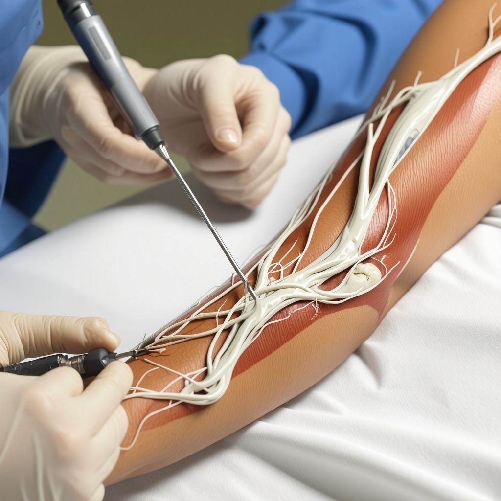
Expert preparation is crucial to optimize test accuracy. Patients should avoid lotions or oils on the skin and inform clinicians of any implanted electrical devices, such as pacemakers. During EMG, fine needle electrodes are inserted into targeted muscles to record activity at rest and during contraction. NCS involves placing surface electrodes to stimulate nerves and record responses. While generally safe, these tests may cause transient discomfort or minor bruising. Orthopedic clinicians must interpret results within the clinical context to avoid false positives or negatives, considering factors like patient age, comorbidities, and medication use.
Advanced Diagnostic Challenges and Interpretative Considerations in Orthopedic Nerve Testing
One complex aspect is differentiating between neuropathic and myopathic patterns, which demands nuanced electrophysiological expertise. Additionally, partial nerve injuries or early-stage neuropathies can present subtle abnormalities that challenge conventional interpretation. Recent advances in EMG and NCS techniques, including high-resolution ultrasound-guided needle placement and quantitative nerve conduction metrics, enhance diagnostic precision. Orthopedic experts must stay abreast of evolving protocols to refine patient assessment and tailor interventions effectively.
What Are the Limitations and Emerging Innovations in EMG and NCS for Orthopedic Applications?
Despite their utility, EMG and NCS have limitations such as discomfort, operator dependency, and reduced sensitivity in detecting proximal nerve lesions. Emerging technologies like microneurography and magnetoneurography hold promise for non-invasive, real-time nerve function assessment but remain largely experimental. Integration with advanced imaging and biomarker analysis may soon redefine diagnostic paradigms in orthopedic nerve disorders.
For those seeking deeper insights into orthopedic nerve testing and advanced spine care, explore expert resources including top orthopedic spine specialists to trust in 2025 and comprehensive guides on effective non-surgical care for herniated discs.
Engage with our orthopedic community by sharing your professional experiences or questions about nerve testing methodologies to foster collective expertise advancement.
According to a detailed review published in the Journal of Clinical Neurophysiology, EMG and NCS remain vital for diagnosing peripheral nerve disorders, emphasizing the importance of operator skill and patient-specific factors in accurate interpretation.
Refining Diagnostic Accuracy: Integrating EMG and NCS with Multimodal Orthopedic Assessments
While EMG and NCS provide crucial electrophysiological data, their diagnostic power is significantly enhanced when combined with advanced imaging techniques such as high-resolution ultrasound and magnetic resonance imaging (MRI). Ultrasound guidance allows precise needle placement during EMG, minimizing patient discomfort and improving signal reliability. MRI complements nerve conduction studies by visualizing structural abnormalities like nerve entrapments and soft tissue lesions that may be contributing to clinical symptoms. Orthopedic specialists increasingly adopt an integrated diagnostic approach to correlate electrophysiological findings with anatomical imaging, thereby improving differential diagnosis and tailoring patient-specific management plans.
Optimizing Patient Outcomes: Tailored Therapeutic Strategies Based on EMG/NCS Findings
Interpretation of EMG and NCS results is pivotal not just for diagnosis but also for guiding therapeutic decisions, including the choice between conservative management and surgical intervention. For instance, electrodiagnostic evidence of severe nerve conduction block or axonal loss may prompt consideration of decompression surgery, whereas milder abnormalities might warrant physical therapy and pharmacologic pain control. Furthermore, serial EMG/NCS testing enables monitoring of nerve regeneration and recovery following treatment, offering objective metrics to adjust rehabilitation protocols effectively. This dynamic utilization underscores the importance of orthopedic specialists’ expertise in electrophysiology for optimizing functional recovery.
How Can Emerging Technologies Revolutionize EMG and NCS Precision in Orthopedic Practice?
Emerging modalities such as high-density surface EMG and automated nerve conduction analysis algorithms present exciting avenues to enhance diagnostic sensitivity and reproducibility. High-density EMG arrays capture spatial muscle activation patterns with unprecedented resolution, facilitating earlier detection of neuropathic changes. Artificial intelligence-driven interpretation tools promise to reduce operator dependency and standardize reporting, addressing one of the main limitations of traditional EMG/NCS. Orthopedic clinicians and researchers are actively exploring these innovations to refine diagnostic workflows and improve patient care outcomes.
For professionals interested in advancing their understanding of cutting-edge orthopedic nerve diagnostics, consider exploring resources like minimally invasive back pain treatments explained and expert guidance on choosing the right orthopedic surgeon for your spine.
We encourage readers to share insights or questions about integrating advanced EMG/NCS techniques in orthopedic practice to foster a collaborative knowledge base within this specialized field.
According to a comprehensive analysis published in Current Opinion in Neurology, advancements such as high-density EMG and AI-based interpretation are poised to transform electrodiagnostic testing, enhancing both diagnostic accuracy and clinical utility in musculoskeletal disorders.
Quantitative Electrodiagnostics: Unlocking New Dimensions in Nerve Function Analysis
The transition from qualitative to quantitative assessment in EMG and NCS embodies a paradigm shift in orthopedic diagnostics. Modern quantitative electrodiagnostics involve detailed numeric measurements of nerve conduction velocities, amplitude ratios, and temporal dispersion, enabling clinicians to detect subtle pathophysiological changes that often elude traditional interpretation. For example, quantitative sensory nerve conduction studies (QSCS) provide objective data on small fiber function, critical in early diabetic neuropathy and chemotherapeutic neurotoxicity evaluations. Incorporating such metrics enhances diagnostic granularity and facilitates longitudinal monitoring with greater sensitivity.
Moreover, advanced signal processing techniques, including wavelet transforms and machine learning classifiers, refine the extraction of diagnostic features from raw EMG signals. These approaches can delineate complex muscle activation patterns, discriminate between neurogenic and myogenic pathologies, and predict prognosis with improved accuracy. Orthopedic practitioners integrating these methodologies can expect a significant elevation in diagnostic confidence, especially in ambiguous clinical scenarios.
How Can Machine Learning Algorithms Improve EMG/NCS Diagnostic Specificity and Reduce Operator Bias?
Machine learning (ML) offers transformative potential by automating pattern recognition and interpretation in EMG and NCS data. Supervised learning models trained on vast datasets can identify nuanced electrophysiological signatures correlating with specific neuropathies or muscular disorders, surpassing human interpretative limits. ML algorithms also mitigate inter-operator variability by standardizing analysis pipelines, thus enhancing reproducibility and reducing diagnostic discrepancies.
Studies have demonstrated that convolutional neural networks (CNNs) can classify EMG signals with high accuracy, facilitating early detection of conditions such as amyotrophic lateral sclerosis (ALS) and peripheral neuropathies. Additionally, ensemble methods combining multiple algorithmic outputs enhance robustness against noise and artifacts common in clinical recordings. Such innovations not only expedite diagnostic workflows but also empower orthopedic clinicians to tailor therapies more precisely based on refined electrophysiological phenotypes (Zhou et al., Frontiers in Neuroscience, 2020).
Integration of High-Resolution Ultrasound with EMG/NCS: A Synergistic Approach to Peripheral Nerve Pathology
High-resolution ultrasound (HRUS) has emerged as an indispensable adjunct to electrodiagnostic testing by enabling real-time visualization of peripheral nerve morphology and surrounding structures. Its non-invasive, dynamic imaging capability complements EMG and NCS by precisely localizing sites of nerve entrapment, inflammation, or traumatic injury. The fusion of HRUS findings with electrophysiological data provides a comprehensive pathoanatomic context, improving preoperative planning and prognostication.
For instance, ultrasound-guided EMG allows clinicians to target deep or anatomically complex muscles with unparalleled accuracy, reducing procedural discomfort and enhancing signal fidelity. Moreover, HRUS can detect nerve swelling, fascicular disruptions, or vascular changes correlating with conduction abnormalities, thereby corroborating or refining electrodiagnostic conclusions.
Such multimodal assessment is particularly valuable in complex cases of entrapment neuropathies like cubital tunnel syndrome or multifocal motor neuropathy, where isolated EMG/NCS findings may be inconclusive. Orthopedic specialists adept in integrating HRUS into their diagnostic algorithm achieve superior patient stratification and individualized management strategies.
Future Directions: Towards Personalized, Predictive Orthopedic Neurodiagnostics
The convergence of bioinformatics, wearable neurotechnology, and telemedicine heralds a new era of personalized orthopedic neurodiagnostics. Emerging wearable EMG sensors and portable nerve conduction devices enable continuous monitoring of neuromuscular function in real-world settings, capturing dynamic fluctuations undetectable in traditional clinical environments. This continuous data stream, analyzed through advanced AI frameworks, promises predictive insights into disease progression and treatment responsiveness.
Additionally, integration with genetic and molecular biomarkers could soon allow orthopedists to stratify patients based on individual susceptibility to nerve injury or regeneration potential, tailoring interventions with unprecedented precision. Collaborative research efforts focusing on multimodal data fusion and real-time analytics are critical to realizing this vision.
For orthopedic clinicians committed to advancing their practice, delving deeper into these cutting-edge developments offers a pathway to elevate patient outcomes and refine diagnostic acumen. Engage with specialized forums and continuous education platforms to remain at the forefront of orthopedic nerve diagnostics innovation.
Decoding Complex Neuropathies: Integrative Strategies Beyond Conventional EMG/NCS
In complex clinical scenarios where conventional EMG and NCS yield equivocal results, orthopedic specialists increasingly turn to integrative diagnostic algorithms encompassing neuroimaging, advanced electrophysiological metrics, and biomolecular profiling. Such multifaceted approaches facilitate discrimination between overlapping neuropathic and myopathic entities, enhancing diagnostic precision. For instance, quantitative EMG parameters combined with diffusion tensor imaging (DTI) of peripheral nerves elucidate microstructural changes underlying chronic compressive neuropathies, guiding targeted interventions.
What cutting-edge electrophysiological biomarkers are emerging to refine nerve injury prognostication in orthopedics?
Recent research highlights novel biomarkers derived from advanced EMG signal decomposition, such as motor unit potential instability indices and high-frequency oscillation analysis, which correlate strongly with nerve regeneration capacity and functional recovery trajectories. These parameters provide prognostic granularity surpassing traditional amplitude and latency measures, enabling personalized rehabilitation planning. Incorporating such biomarkers into routine electrodiagnostic protocols requires sophisticated signal processing tools and specialized clinician training.
Moreover, the integration of microneurography techniques allows direct recording of single afferent nerve fiber activity, offering unprecedented insights into nociceptive and autonomic nerve function in musculoskeletal disorders. This modality complements EMG/NCS by elucidating small fiber involvement often missed in standard testing, informing nuanced therapeutic strategies.
Leveraging Artificial Intelligence and Deep Learning for Automated EMG/NCS Interpretation
Artificial intelligence (AI), particularly deep learning architectures, has revolutionized the interpretation of electrodiagnostic data by automating pattern recognition and reducing inter- and intra-operator variability. Convolutional neural networks (CNNs) trained on extensive annotated EMG and NCS datasets can accurately classify neuropathic versus myopathic signatures and detect subtle demyelination patterns. This automation accelerates diagnostic workflows, enhances reproducibility, and aids in early detection of subclinical nerve pathologies.
Furthermore, AI-driven decision support systems integrate electrodiagnostic findings with clinical and imaging data, offering comprehensive diagnostic impressions and prognostic predictions. These systems harness large-scale multimodal datasets and continuously learn from new cases, embodying a dynamic, evidence-based diagnostic paradigm. Orthopedic clinicians adopting such technologies can expect improved accuracy and efficiency in managing peripheral nerve disorders.
Precision Medicine Meets Orthopedic Electrodiagnostics: Toward Personalized Nerve Disorder Management
The convergence of quantitative electrodiagnostics, molecular biomarkers, and patient-specific genetic profiles heralds a new era of precision medicine in orthopedic nerve diagnostics. Tailoring interventions based on individual electrophysiological phenotypes and genetic susceptibilities optimizes therapeutic efficacy and minimizes adverse outcomes. For example, pharmacogenomic data combined with EMG/NCS metrics can guide personalized neuropathic pain management strategies, enhancing patient quality of life.
Additionally, wearable biosensors capable of continuous EMG monitoring empower real-time assessment of neuromuscular dynamics during rehabilitation, facilitating adaptive therapy adjustments. Such technologies exemplify the shift toward proactive, data-driven orthopedic care.
For an in-depth exploration of these advancements and expert guidance on integrating AI and quantitative electrodiagnostics into clinical practice, consult resources like the Journal of Clinical Neurophysiology and the Current Opinion in Neurology.
Engage with our advanced orthopedic forum to discuss these innovations and share your clinical experiences, contributing to the collective expertise in neuromuscular diagnostics.
Bridging Diagnostic Modalities: The Synergistic Role of High-Resolution Ultrasound and Electrodiagnostics
High-resolution ultrasound (HRUS) not only enhances anatomical localization but also facilitates dynamic assessment of nerve mobility and vascularity, critical in diagnosing complex entrapment syndromes. When combined with AI-enhanced EMG/NCS data, HRUS provides a multidimensional diagnostic perspective that refines surgical planning and prognostic evaluation.

Expert Insights & Advanced Considerations
Precision in Electrodiagnostic Interpretation Requires Multimodal Integration
Orthopedic practitioners must embrace a holistic diagnostic approach, combining EMG and NCS with high-resolution ultrasound and MRI. This synergy enhances localization accuracy of nerve pathologies and refines clinical decision-making beyond isolated electrophysiological data.
Quantitative Metrics and Machine Learning Are Reshaping Diagnostic Sensitivity
The transition from qualitative to quantitative electrodiagnostics, powered by AI algorithms, is elevating the detection of subtle neuropathies and myopathies. Machine learning reduces operator bias, standardizes interpretation, and facilitates earlier intervention strategies tailored to individual patient profiles.
Emerging Biomarkers from Advanced Signal Analysis Offer Prognostic Value
Novel electrophysiological biomarkers such as motor unit potential instability indices and high-frequency oscillation parameters provide deeper insight into nerve regeneration potential. Incorporating these into routine protocols empowers personalized rehabilitation and more accurate prognostication in orthopedic nerve disorders.
Wearable Neurotechnology and Telemedicine Are Pioneering Personalized Monitoring
Continuous neuromuscular monitoring through wearable EMG sensors integrated with telemedicine platforms allows dynamic assessment of nerve function in real-life settings. This real-time data stream supports predictive analytics and adaptive treatment modifications, heralding a new paradigm in orthopedic neurodiagnostics.
Curated Expert Resources
Journal of Clinical Neurophysiology: Offers in-depth reviews and cutting-edge research on electrodiagnostic techniques, essential for clinicians seeking authoritative guidance on EMG and NCS advancements.
Top Orthopedic Spine Specialists to Trust in 2025: A curated resource highlighting leading experts who exemplify best practices in integrating advanced nerve diagnostics in orthopedic care.
Current Opinion in Neurology: Provides comprehensive analyses of emerging technologies such as high-density EMG and AI-driven interpretation methods, crucial for staying abreast of evolving diagnostic paradigms.
Minimally Invasive Back Pain Treatments Explained: Details innovative therapeutic approaches informed by precise electrodiagnostic assessments, offering practical strategies for clinicians.
Choosing the Right Orthopedic Surgeon for Your Spine: Guidance on selecting specialists who prioritize advanced diagnostic modalities like EMG and NCS to optimize patient outcomes.
Final Expert Perspective
The integration of EMG and NCS within modern orthopedic diagnostics represents a pivotal advancement in neuromuscular evaluation. By coupling electrophysiological insights with imaging, quantitative analytics, and AI-assisted interpretation, orthopedic specialists can achieve unprecedented diagnostic precision and personalized treatment planning. Embracing wearable technologies and emerging biomarkers further extends the capacity to monitor and predict nerve recovery dynamically. For clinicians striving to elevate their practice and improve patient outcomes, engaging deeply with these innovations is indispensable. We invite you to explore advanced resources and share your professional insights to collectively advance the state of orthopedic nerve diagnostics. To connect with expert orthopedic care or learn more about minimally invasive approaches, visit our contact page or explore detailed guides such as minimally invasive back pain treatments explained.