My First Encounter with Diagnostic Imaging — A Personal Story
Not long ago, I experienced a persistent back pain that just wouldn’t go away. After trying various remedies, I decided to consult a trusted orthopedic specialist. During my visit, I learned that diagnostic imaging plays a crucial role in identifying the root cause of spine and back pain. This personal experience made me realize how vital these imaging techniques are in modern orthopedics.
Understanding the Different Types of Diagnostic Imaging
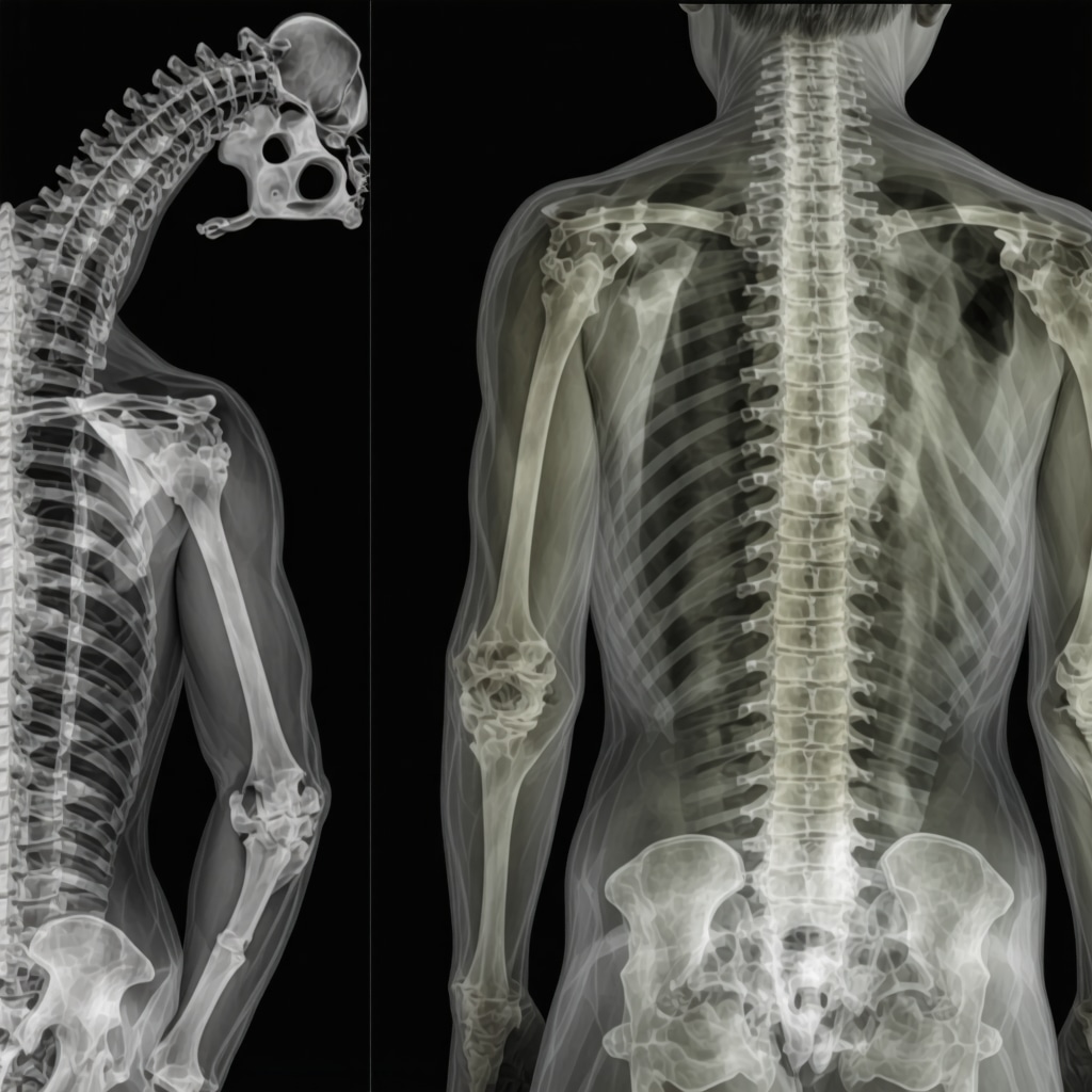
As someone curious about medical procedures, I was eager to understand the various imaging modalities. I discovered that X-rays are often the first step, providing clear images of bones and structural abnormalities. MRI scans, on the other hand, offer detailed views of soft tissues, discs, and nerves, which are essential in diagnosing herniated discs or ligament injuries. CT scans serve as a bridge between the two, giving comprehensive cross-sectional images.
The Role of MRI in Diagnosing Back Pain
My doctor explained that MRI is particularly effective in detecting issues like nerve compression or disc degeneration. I was impressed by how detailed these images are, enabling precise treatment planning. For instance, if surgery becomes necessary, MRI findings help surgeons determine the best approach, whether minimally invasive or more traditional methods. I also learned that MRI doesn’t use ionizing radiation, making it safer for patients.
How Do Imaging Results Shape Treatment Plans?
Seeing my own MRI images was eye-opening. It was clear how these images guide the treatment process—from conservative care like physical therapy to advanced surgical interventions. For example, if facet joint pain is diagnosed, options like targeted injections or radiofrequency ablation might be considered, as discussed in authoritative sources like the facet joint treatment guide. These imaging results give both patients and physicians a roadmap for effective management.
What Advances Are Making Diagnostic Imaging Safer and More Accurate?
In my research, I found that recent advancements, such as high-resolution MRI and low-dose CT scans, are improving diagnostic accuracy while minimizing risks. Experts in the field continually develop new protocols to enhance patient safety and image clarity. For those interested, exploring the latest imaging techniques can be very enlightening—more information can be found on reputable medical websites or through trusted orthopedic specialists.
If you’re navigating similar issues, I encourage you to discuss these imaging options thoroughly with your healthcare provider. Sharing experiences and asking questions can help you make informed decisions about your spine health. Feel free to leave a comment or reach out for more insights on effective back pain management and diagnostic strategies in orthopedics.
Remember, understanding your diagnostic options empowers you to take control of your health journey. For additional guidance, consider visiting trusted sources like the top spine specialists list.
How Are Emerging Technologies Transforming Orthopedic Diagnostic Imaging?
Recent breakthroughs in imaging technology are revolutionizing how orthopedic specialists diagnose and treat spine and joint conditions. High-field MRI machines now offer unprecedented detail, enabling clinicians to detect subtle soft tissue injuries and early degenerative changes that were previously difficult to visualize. Additionally, AI-enhanced imaging algorithms are assisting radiologists in interpreting scans more accurately and swiftly, reducing diagnostic errors and expediting treatment decisions.
One promising development is the advent of 3D imaging techniques, which provide comprehensive spatial views of complex anatomical structures. These advancements facilitate precise surgical planning, particularly in minimally invasive procedures. For example, 3D reconstructions of herniated discs or facet joint problems enable surgeons to target pathology more effectively, potentially improving outcomes and reducing recovery time.
Could These Innovations Lead to Safer, More Efficient Diagnoses in Orthopedics?
Absolutely. Innovations like low-dose CT scans are making it possible to obtain high-quality images with minimal radiation exposure—an important consideration for patient safety. Furthermore, the integration of functional imaging techniques, such as diffusion tensor imaging (DTI), is opening new doors for understanding nerve injuries and degeneration. These tools provide insights into nerve integrity and help tailor personalized treatment plans, from conservative management to surgical intervention.
Experts emphasize that staying abreast of these technological advancements is vital for orthopedic practitioners aiming to deliver top-tier care. Resources like the latest advances in orthopedic imaging offer valuable updates on cutting-edge techniques.
What Are the Practical Implications for Patients and Practitioners?
For patients, these innovations mean faster, more accurate diagnoses that can lead to earlier interventions and better prognosis. For practitioners, it involves adopting new protocols, investing in advanced imaging equipment, and continuously updating their knowledge base. Proper interpretation of these images is crucial; thus, collaboration with radiologists and specialists is essential to maximize diagnostic precision.
Furthermore, emerging imaging modalities are helping to monitor treatment progress more effectively. For example, serial MRI scans can assess the healing of disc herniations or post-surgical recovery, guiding rehabilitation and activity recommendations. This dynamic assessment capability ensures that care remains aligned with the patient’s evolving condition.
If you’re interested in exploring more about how these innovations are shaping orthopedic care, I recommend reviewing trusted sources or consulting with experienced specialists. Sharing your questions and experiences can foster a deeper understanding and improve your health outcomes. Feel free to comment below or reach out to your healthcare provider for personalized advice.
Remember, understanding the latest diagnostic tools empowers you to make informed decisions about your spine and joint health. For insights into expert care options, visit the top spine specialists in 2025.
Embracing the Next Generation of Imaging Technologies — My Personal Reflections
As I continue to explore the evolving landscape of diagnostic imaging, I realize that each new advancement not only enhances our diagnostic precision but also reshapes how we approach patient care. From my own journey through understanding MRI and CT scans to delving into cutting-edge innovations, I’ve come to appreciate the profound impact these technologies have on treatment outcomes.
How Do Personal Experiences Inform Our Expectations of Imaging Advances?
Reflecting on my encounters with imaging, I notice that the stories of other patients often mirror my own—initial uncertainty, followed by reassurance when clear images lead to effective treatment plans. Personally, I find that the integration of AI and 3D imaging not only expedites diagnoses but also offers a more comprehensive view of complex anatomical structures. These tools are gradually transforming the patient experience, making it more transparent and empowering.
What Are the Nuances of Emerging Technologies in Orthopedic Imaging?
One area that fascinates me is the development of high-field MRI machines, which provide exquisite detail that was previously unattainable. This allows clinicians to detect subtle soft tissue injuries and early degenerative changes with greater confidence. Additionally, the advent of functional imaging techniques, such as diffusion tensor imaging (DTI), opens new frontiers in understanding nerve injuries and degeneration—areas that directly influence how we manage conditions like radiculopathy or peripheral neuropathy. For those interested, exploring reputable sources like the latest advances in orthopedic imaging can deepen your understanding of these breakthroughs.
Personally, I believe these innovations will lead to more personalized care, tailored precisely to the patient’s unique pathology, ultimately improving prognosis and quality of life.
Are There Ethical or Practical Challenges in Integrating New Imaging Modalities?
While I am excited about technological progress, I am also aware of challenges such as cost, accessibility, and the need for ongoing training. For instance, high-resolution MRIs and AI-driven interpretation tools require significant investment, which might not be feasible everywhere. Moreover, ensuring that practitioners are adequately trained to interpret these advanced images is crucial to prevent misdiagnosis. These considerations remind me that technological advancement must go hand-in-hand with thoughtful implementation and equitable access.
For patients navigating these options, I suggest discussing with your healthcare provider not only the available imaging modalities but also their implications. Sharing your experiences and questions can foster a more informed and collaborative approach to your care. Feel free to leave comments or reach out for insights on how emerging imaging techniques might benefit your specific condition.
How Can Staying Informed About Imaging Innovations Empower Patients?
Staying updated on technological trends equips you with the knowledge to advocate effectively for your health. It also helps set realistic expectations about diagnosis and treatment timelines. For instance, understanding the benefits of low-dose CT scans can alleviate concerns about radiation exposure, while familiarity with 3D reconstructions can reassure you about surgical planning accuracy. To explore more, visit trusted sources like the top spine specialists in 2025.
In my journey, I’ve learned that embracing these innovations with a critical yet optimistic mindset allows us to navigate the complexities of modern medicine more confidently. As the field continues to evolve, I look forward to seeing how these tools will further refine our approach to diagnosing and treating orthopedic conditions, ultimately leading to better outcomes for all.
Deepening the Personal Connection to Imaging Innovations
My ongoing journey through the landscape of diagnostic imaging has been both enlightening and inspiring. Having first encountered MRI and CT scans during my own back pain ordeal, I became acutely aware of how these technologies not only diagnose but also shape our expectations for treatment outcomes. As I delved deeper, I discovered that the integration of advanced imaging techniques, such as 7 Tesla MRI machines, is opening new frontiers in soft tissue visualization and early degenerative change detection, which were once beyond reach. These innovations are transforming the diagnostic paradigm from reactive to proactive care, allowing for interventions at stages where they can be most effective.
The Nuanced Role of AI and 3D Imaging in Personalized Treatment
One of the most compelling developments I’ve explored is the synergy between artificial intelligence (AI) and 3D imaging reconstruction. AI algorithms, trained on vast datasets, assist radiologists in identifying subtle anomalies with remarkable accuracy, reducing human error and expediting diagnosis. Meanwhile, 3D reconstructions of complex spinal anatomy facilitate surgical planning with unprecedented precision. This combination empowers surgeons to execute minimally invasive procedures with confidence, ultimately improving patient outcomes and reducing recovery times. According to a study published in Nature Medicine, AI-enhanced imaging has demonstrated a significant reduction in diagnostic discrepancies, underscoring its potential to revolutionize orthopedic diagnostics.
Addressing Ethical and Practical Challenges in Technological Adoption
While the promise of these emerging technologies is undeniable, I remain mindful of the ethical and practical hurdles they pose. The high costs associated with deploying high-resolution MRI systems and AI-driven tools can create disparities in access, especially in under-resourced communities. Additionally, the need for specialized training to interpret complex images is a barrier that requires concerted efforts in medical education. Ensuring data privacy and security in AI applications is another critical aspect that demands rigorous standards. Navigating these challenges calls for a balanced approach—one that fosters innovation while promoting equitable healthcare delivery. For those interested in how these issues are being addressed, the latest guidelines from organizations such as the American College of Radiology provide valuable insights.
Empowering Patients through Knowledge and Engagement
From my perspective, informed patients are empowered patients. Staying abreast of technological advancements enables you to actively participate in your care decisions. For example, understanding the benefits of low-dose CT scans or the potential of functional imaging can alleviate fears and foster trust in the diagnostic process. I encourage readers to engage with their healthcare providers about emerging imaging options, ask about the safety and efficacy of new modalities, and seek second opinions when necessary. Sharing your experiences and questions not only enhances your understanding but also contributes to a broader dialogue aimed at improving care standards. To explore more about how advanced imaging can benefit your specific condition, visit trusted sources like the top spine specialists list.
Future Directions: From Research to Routine Practice
Looking ahead, I believe that the convergence of technological innovation, personalized medicine, and ethical stewardship will define the next era of orthopedic diagnostics. The ongoing development of portable, point-of-care imaging devices promises to bring high-quality diagnostics closer to patients, reducing delays and enhancing early intervention. Moreover, as machine learning algorithms become more sophisticated, their integration into routine clinical workflows will likely become seamless, providing real-time diagnostic support. These advancements will not only improve accuracy but also democratize access to expert-level care. For those eager to stay at the forefront of these changes, I recommend following updates from leading research institutions and participating in specialist forums. Your proactive engagement can be a vital component in shaping a future where diagnostic excellence is accessible to all.
Things I Wish I Knew Earlier (or You Might Find Surprising)
The Power of Soft Tissue Imaging
When I first heard about MRI scans, I thought they only showed bones. Turns out, they reveal soft tissues like muscles, ligaments, and nerves in incredible detail. This was a game-changer for my understanding of what’s happening inside the body and how complex injuries can be.
The Safety of MRI Over X-rays
Initially, I was worried about radiation from X-rays and CT scans. Learning that MRI doesn’t use ionizing radiation made me feel more comfortable, especially for repeated imaging or vulnerable populations. It’s reassuring to know that safer options exist without sacrificing diagnostic quality.
The Role of Advanced Technology in Early Detection
Discovering that high-resolution MRI and AI-powered imaging can spot subtle issues early on amazed me. This proactive approach allows for interventions before symptoms become severe, potentially saving patients from more invasive treatments later.
Imaging as a Roadmap for Surgery
Seeing how detailed images guide surgical planning was eye-opening. Precise imaging results mean less invasive procedures, shorter recovery times, and better outcomes. It’s like having a GPS for the surgeon’s tools.
The Future: 3D and Functional Imaging
The development of 3D reconstructions and functional imaging techniques like DTI is fascinating. These tools provide a comprehensive picture, helping tailor treatments uniquely suited to each patient’s condition and anatomy. It’s personalized medicine in action.
Resources I’ve Come to Trust Over Time
- American College of Radiology (ACR): Their guidelines and updates are clear and trustworthy, helping me understand the latest standards in imaging safety and quality.
- National Institutes of Health (NIH): A treasure trove of research articles and breakthroughs, making complex topics accessible and credible.
- PubMed Central: For in-depth studies on new imaging technologies and their clinical benefits, I often turn here for the latest evidence-based info.
- Radiology Masterclass: An excellent resource for learning about different imaging modalities and their practical applications in orthopedics.
Parting Thoughts from My Perspective
Reflecting on my journey with diagnostic imaging, I realize how much these technologies have transformed patient care, including my own understanding of health. They provide a clearer picture, reduce uncertainty, and enable more effective treatments. If you’re navigating similar issues, I encourage you to be proactive in discussing these options with your healthcare provider. Staying informed empowers you to make better decisions and advocate for your health. If this resonated with you, I’d love to hear your thoughts. Feel free to share your experiences in the comments or reach out through your local orthopedic specialist’s office. Remember, understanding your diagnostic options is a vital step toward a healthier, more confident life.
Reading about your personal journey with diagnostic imaging really resonated with me. I recently had a similar experience after enduring persistent neck pain, and I was amazed at how detailed MRI and CT scans provided clarity that traditional exams couldn’t offer. It made a huge difference in getting an accurate diagnosis and tailored treatment plan. I agree that advancements like 3D imaging and AI are truly transformative, especially in reducing diagnostic errors and improving surgical precision. My question is, how do you think these emerging technologies will impact the cost and accessibility of imaging services in the future? I worry that while they benefit patients with access, they might widen the gap for those in under-resourced areas. I’d love to hear your thoughts or experiences with navigating such disparities. Overall, staying informed about these innovations empowers us to make better health decisions and advocate for necessary care, which I find extremely encouraging.