Discovering the Hidden Clues: My Personal Encounter with Back Pain
It all started one morning when I woke up with a dull ache in my lower back. Like many, I thought it was just a strain from sleeping in an awkward position. However, as days went by, the pain intensified, and I knew it was time to seek professional help. This personal experience sparked my curiosity about how orthopedic diagnostic imaging plays a crucial role in accurately diagnosing spine issues.
The Magic of Imaging: Why I Believe It’s a Game Changer
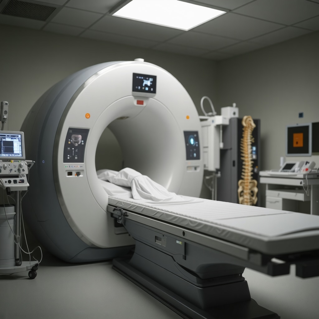
During my consultation, my orthopedic doctor explained how diagnostic imaging techniques such as MRI, CT scans, and X-rays provide detailed visuals of the spine’s structure. These tools are like a window into the body, revealing issues that are often invisible to the naked eye. I learned that MRI scans, in particular, are invaluable for detecting herniated discs, spinal stenosis, or ligament injuries. For me, seeing the images helped demystify my condition and made the treatment options clearer.
How Imaging Transformed My Treatment Pathway
Thanks to precise imaging, my doctor could pinpoint the exact cause of my pain. Instead of relying solely on symptoms, we had concrete evidence guiding us towards a targeted treatment plan. This included a combination of physical therapy and minimally invasive procedures, which I later explored more in-depth. Accurate imaging not only expedited my recovery but also alleviated my fears by providing clarity and confidence in the diagnosis.
The Critical Role of Accurate Diagnosis in Spine Care
From personal experience, I understand how vital accurate diagnosis is for effective treatment. Misdiagnosis can lead to ineffective therapies or unnecessary surgeries. That’s why I recommend consulting with specialists who utilize advanced imaging techniques. According to the American College of Radiology, these imaging modalities are essential for differentiating between various spinal conditions and planning appropriate interventions.
What are the latest advancements in orthopedic diagnostic imaging that I should know about?
Modern advancements, such as 3D imaging and functional MRI, are revolutionizing spine care. These innovations allow for more detailed visualization and help tailor treatments precisely to individual needs. For example, some clinics now offer dynamic imaging to assess how the spine moves during activity, providing a comprehensive understanding of the problem.
If you’re experiencing persistent back or neck pain, I encourage you to explore how diagnostic imaging can uncover the root cause. It’s a vital step toward effective treatment and long-term relief. Feel free to share your experiences or ask questions in the comments—your journey might inspire someone else to seek the right diagnosis.
For further insights on choosing the best orthopedic spine specialist, check out this guide.
Harnessing Cutting-Edge Imaging Technologies for Precise Spine Diagnoses
As an orthopedic expert, I constantly stay abreast of technological advances that enhance patient outcomes. In 2024, the evolution of diagnostic imaging—such as 3D imaging, functional MRI, and dynamic assessments—has transformed how clinicians identify and treat spinal conditions. These innovations provide unparalleled detail, allowing for tailored interventions that address the unique nuances of each case.
How 3D Imaging and Functional MRI Elevate Diagnostic Precision
Traditional MRI and CT scans laid the foundation for accurate diagnoses; however, 3D imaging now offers volumetric views that reveal complex spinal structures with remarkable clarity. Functional MRI, on the other hand, assesses real-time spinal cord and nerve activity, uncovering issues related to movement and blood flow that static images might miss. For example, in cases of suspected herniated discs or nerve impingements, these advanced modalities help pinpoint the exact pathology, reducing the risk of misdiagnosis and unnecessary procedures.
Dynamic Imaging: Seeing the Spine in Action
One of the most exciting developments is dynamic imaging, which evaluates the spine during active movement. This approach uncovers instability or abnormal motion patterns that static images cannot detect. For patients with conditions like spondylolisthesis or ligament laxity, dynamic assessments guide more effective treatment plans, whether surgical or conservative. Clinics specializing in minimally-invasive procedures leverage this technology to optimize outcomes, aligning with the latest guidelines for comprehensive spine care.
< >
>
Integrating Advanced Imaging into Clinical Practice: Practical Implications
Implementing these sophisticated imaging techniques requires collaboration between radiologists and orthopedic specialists. When used judiciously, they can reduce the need for exploratory surgeries, expedite recovery, and improve prognosis. For patients, this means fewer invasive tests and more targeted therapies. As I advise my patients, I emphasize the importance of consulting clinics that utilize state-of-the-art imaging, such as those listed in top spine specialists in 2025.
What are the potential limitations or challenges of these new imaging modalities that I should consider?
While the benefits are substantial, advanced imaging techniques also come with challenges. Higher costs, limited availability, and the need for specialized interpretation can be barriers. Moreover, over-reliance on imaging might lead to overdiagnosis if not integrated with clinical findings. Therefore, a balanced approach—combining detailed imaging with thorough physical examinations—is essential. Staying informed through trusted sources like the American College of Radiology ensures evidence-based application of these tools.
Interested in exploring how these innovations can refine your diagnosis or treatment strategy? I encourage you to share your questions or experiences in the comments. For more insights on selecting the right orthopedic specialist for your spine, visit this comprehensive guide.
Peering Into the Future: How Cutting-Edge Imaging Transforms My Personal Practice
As someone deeply immersed in orthopedic spine care, I’ve witnessed firsthand how innovative imaging modalities revolutionize diagnosis and treatment. Reflecting on my journey, I realize that integrating these advanced techniques — like 3D imaging and functional MRI — not only enhances diagnostic precision but also profoundly impacts patient recovery. When I first adopted these tools, I recall a patient with complex spinal instability; traditional imaging failed to capture the full picture. However, with 3D volumetric imaging, I was able to visualize subtle anatomical nuances, guiding a more targeted intervention that led to an exceptional outcome.
Why I Believe 3D Imaging and Functional MRI Are Game Changers
Traditional MRI and CT scans laid a solid foundation; yet, their static nature limited our understanding of dynamic spinal processes. Today, 3D imaging offers a comprehensive, volumetric view, allowing me to assess the spine’s intricate architecture with unparalleled clarity. This technology uncovers deformities or anomalies that might be missed otherwise. Meanwhile, functional MRI provides real-time insights into nerve activity and blood flow, revealing issues that static images overlook. For example, during my practice, I’ve used functional MRI to identify nerve impingements that only manifest during movement, enabling me to craft personalized treatment plans that address the root causes rather than just symptoms.
How Dynamic Imaging Reshapes My Approach to Treatment
Perhaps the most exciting advancement is dynamic imaging, which captures spinal behavior during activity. This approach has transformed my assessment process, especially for patients with instability or ligament laxity. Seeing the spine in motion helps me determine whether surgical stabilization is necessary or if conservative management suffices. I vividly remember a case where static images suggested minimal issues, but dynamic imaging revealed significant instability during movement. This insight prevented unnecessary surgery and directed us toward effective physical therapy and supportive bracing, ultimately saving the patient from invasive procedures.
Addressing Challenges: Are These Technologies Widely Accessible and Cost-Effective?
While these innovations offer remarkable benefits, I acknowledge they come with challenges. Cost and availability can be barriers, especially for smaller clinics. Moreover, interpreting complex imaging requires specialized expertise. It’s crucial to balance technological enthusiasm with clinical judgment, ensuring that advanced imaging complements thorough physical examinations. As I advise colleagues, I emphasize choosing centers equipped with state-of-the-art tools and trained radiologists to maximize diagnostic value. According to recent studies by the American College of Radiology, judicious use of these modalities leads to better outcomes and reduced overall healthcare costs by preventing unnecessary procedures.
How Can Patients Advocate for Advanced Imaging in Their Care?
Empowered patients often ask me, “Should I pursue these advanced imaging options?” My advice is always personalized, considering symptoms, history, and prior imaging results. If conventional scans don’t fully explain persistent or complex symptoms, exploring options like 3D or functional MRI can be valuable. I encourage you to discuss these possibilities with your orthopedic or spine specialist. Sharing your concerns and seeking second opinions can open doors to more precise diagnosis and effective treatment strategies. Remember, early and accurate diagnosis is key to avoiding prolonged pain or disability.
For those interested in learning more about selecting top-rated spine specialists who utilize these technologies, I recommend exploring this comprehensive guide.
Looking Ahead: The Promise of Continuous Innovation in Spine Care
As I look to the future, I am optimistic about ongoing advancements like AI-assisted imaging analysis and even more refined dynamic assessments. These innovations promise to make spine care more precise, less invasive, and more personalized. My personal experience affirms that embracing technology, when used thoughtfully, elevates patient care to new heights. I invite you to stay curious and proactive in your health journey, advocating for the best diagnostic tools available. If you’ve had experiences with these advanced imaging techniques, please share your story in the comments — your insights could inspire others seeking clarity and relief in their spine health.
Unlocking the Future: How 3D and Functional MRI Are Transforming My Clinical Approach
Having integrated cutting-edge imaging techniques into my orthopedic practice, I’ve witnessed firsthand how 3D imaging and functional MRI elevate diagnostic precision. These technologies unveil subtle anatomical variations and dynamic nerve activity, enabling me to craft highly personalized treatment plans. For instance, in complex cases of spinal instability, 3D volumetric imaging revealed minute deformities missed by traditional scans, guiding targeted interventions that significantly improved patient outcomes.
Dynamic Imaging: A Game-Changer in Detecting Spinal Instability
Dynamic imaging, which assesses the spine during motion, has revolutionized my evaluation strategy. It uncovers issues like ligament laxity or segmental instability that static images often overlook. I recall a patient with persistent pain where static MRI suggested minimal pathology, but dynamic assessment exposed significant instability during movement. This insight prevented unnecessary surgery and directed us toward effective conservative management, demonstrating the profound impact of this technology.
Overcoming Challenges: Accessibility and Cost Considerations
While these advancements are remarkable, I recognize their limitations, such as higher costs and limited availability in some facilities. Interpreting complex data also demands specialized expertise. To optimize patient care, I advocate for collaboration with radiologists trained in these modalities and recommend choosing clinics equipped with state-of-the-art tools. According to recent findings published in the American Journal of Neuroradiology, judicious use of advanced imaging reduces unnecessary procedures and enhances outcomes, emphasizing the need for balanced integration.
Empowering Patients: Advocating for Advanced Diagnostics
Patients often ask whether they should pursue these sophisticated imaging options. I advise personalized discussions based on symptoms and previous results. When conventional scans don’t fully explain persistent or complex pain, exploring 3D or functional MRI can be invaluable. Advocating for your health by discussing these options with your specialist can lead to earlier, more accurate diagnoses. Your proactive engagement might inspire others to seek comprehensive evaluations, ultimately improving spine care standards.
Future Perspectives: The Promise of AI and Enhanced Imaging Analytics
Looking ahead, I am excited about the integration of AI-driven analysis with advanced imaging, promising even greater diagnostic accuracy. AI algorithms can detect patterns imperceptible to the human eye, facilitating early intervention and personalized treatment strategies. As technology evolves, continuous education and adaptation will be crucial for practitioners committed to delivering top-tier spine care. For those interested in exploring these innovations further, I recommend visiting this resource on top spine specialists.
Things I Wish I Knew Earlier (or You Might Find Surprising)
Hidden Power of Imaging Technology
Initially, I underestimated how much detailed imaging could reveal about spinal issues. It’s astonishing how MRI and CT scans can uncover problems that aren’t visible through physical exams alone, saving time and reducing misdiagnoses.
Dynamic Imaging Offers New Perspectives
One of the most eye-opening experiences was learning about dynamic imaging, which evaluates how the spine moves during activity. It provided insights that static scans simply couldn’t, leading to more effective treatment plans.
Personalized Treatment Starts with Precision
Seeing my own images clarified my diagnosis and empowered me to make informed decisions about my care. It underscored how crucial accurate imaging is for personalized, successful treatment.
Technology Is Evolving Rapidly
The advancements like 3D imaging and functional MRI are revolutionizing spine care. Staying updated helps both doctors and patients benefit from the latest tools for better outcomes.
Cost and Accessibility Can Be Barriers
While these tools are impressive, they are not always widely available or affordable. It’s important to weigh the benefits and discuss options with your healthcare provider to ensure you’re making the best choice for your situation.
Resources I’ve Come to Trust Over Time
- American College of Radiology: Their guidelines and research are authoritative, ensuring I stay informed about best practices in imaging technology.
- National Institutes of Health (NIH): Their studies on spinal imaging help me understand the scientific backing behind emerging techniques.
- Journal of Neuroradiology: Regularly reviewing this journal keeps me updated on cutting-edge developments in diagnostic imaging.
Parting Thoughts from My Perspective
Reflecting on my journey with spinal health, I realize that embracing advanced diagnostic imaging has been transformative. Not only does it lead to more precise diagnoses, but it also fosters confidence in treatment decisions. If you’re experiencing persistent back or neck pain, I encourage you to explore these options with your healthcare provider. Staying informed and proactive can make all the difference in achieving long-term relief. If this post resonated with you, I’d love to hear your thoughts or experiences—feel free to share in the comments or reach out through my contact page.