My Unexpected Journey Through Back Pain Imaging
When I first experienced persistent lower back pain, the thought of undergoing medical imaging was daunting. I remember my initial doctor’s recommendation was to start with an X-ray, which seemed like the simplest option. But having done some personal research, and from my experience visiting orthopedic specialists, I wondered if MRI might be a better first step. This personal exploration led me to understand the distinct advantages and limitations between MRI and X-ray as back pain imaging options.
Why I Leaned Towards MRI for My Back Pain Diagnosis
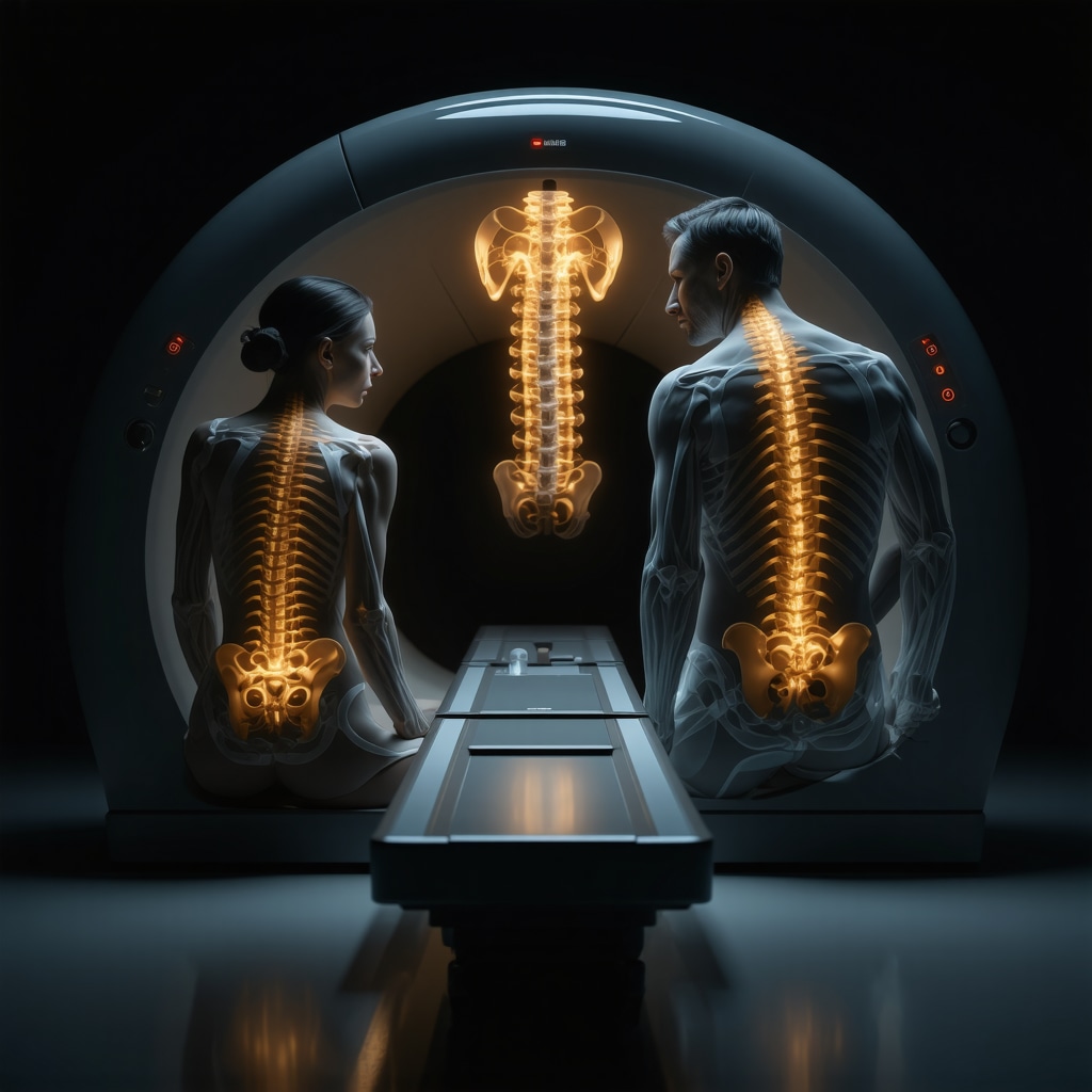
X-rays have always been the go-to for bone injuries or fractures, but I learned they provide limited insight when it comes to soft tissues like discs, nerves, and muscles. My back pain was more than just a twinge—it felt like something deeper, possibly related to disc or nerve issues. MRI, or Magnetic Resonance Imaging, offers detailed images of both bones and soft tissues without radiation exposure. I found that MRI’s ability to visualize herniated discs, nerve impingements, and inflammation gave me a clearer understanding of my condition.
Of course, MRI isn’t perfect: it’s more expensive and not always the immediate first choice if the symptoms are straightforward. However, for complex or chronic back pain, many orthopedic spine specialists advocate for MRI as the first imaging modality. In fact, according to the American Academy of Orthopaedic Surgeons, MRI is often preferred when neurological symptoms suggest nerve involvement (AAOS Back Pain Resource).
How do I decide between MRI and X-Ray for back pain imaging?
That was the question I faced and I want to share what helped me decide. If pain is recent, mild, and without neurological symptoms, an X-ray might be sufficient to rule out fractures or arthritis. But if symptoms include numbness, tingling, weakness, or shooting pain down the leg, MRI is often the smarter first choice. I recommend consulting a trusted spine doctor to evaluate your symptoms and guide imaging decisions. If you want to explore expert advice, check out this helpful guide for choosing the right spine doctor in NJ (choosing the right spine doctor).
Lessons From My Imaging Experience and What You Should Know
After undergoing an MRI, the detailed images revealed a mild herniated disc pressing against a nerve root. This diagnosis was missed on my X-ray, which only showed some mild degenerative changes. This experience taught me that sometimes starting with an MRI can save time, reduce repeated imaging, and lead to more targeted treatment earlier.
If you’re dealing with back pain, I encourage you to explore all your options. And if you’re interested in effective non-surgical care for disc issues, I found valuable insights in this article on non-surgical herniated disc treatments.
Have you ever faced a similar dilemma about imaging for back pain? I’d love to hear your story or questions in the comments below. Sharing our experiences can help many of us navigate this confusing process with more confidence.
Beyond the Basics: When Advanced Imaging Becomes Essential
While X-rays and MRIs cover much ground, sometimes the complexity of back pain calls for even more specialized imaging techniques. For instance, CT scans can provide detailed views of bone structures and are often used when an MRI is contraindicated or unavailable. Meanwhile, newer modalities like functional MRI and diffusion tensor imaging are emerging tools that might one day offer enhanced insights into nerve integrity and soft tissue health. Understanding these options can empower patients to engage more knowledgeably with their orthopedic specialists.
What factors should influence the choice of imaging modality in complex spinal cases?
Deciding which imaging tool to use involves balancing several variables: the nature and duration of symptoms, prior imaging results, patient medical history, and potential contraindications to certain tests (such as metal implants precluding MRI). For example, in cases of persistent radiculopathy where initial MRI findings are inconclusive, a CT myelogram might be recommended to better visualize nerve root compression. Additionally, some patients may require repeat imaging to monitor progression or response to treatment, which has implications for radiation exposure and cost.
Consultation with an experienced orthopedic spine specialist is crucial to tailor imaging strategies appropriately. For those interested in learning how to choose the best spine doctor in New Jersey, this comprehensive guide offers practical advice.
Insurance and Imaging: Navigating Authorization and Coverage Challenges
One often overlooked aspect is insurance authorization for advanced imaging like MRI or CT scans. Many patients face delays or denials, which can prolong diagnosis and treatment. Understanding how to effectively obtain prior authorization can facilitate timely care. Experienced orthopedic clinics often assist patients through this process, reducing administrative burdens and ensuring appropriate imaging is performed.
For more detailed tips on expediting insurance approvals, see orthopedic prior authorization tips.
Integrating Imaging Findings Into Personalized Treatment Plans
Imaging results are only as valuable as the treatment decisions they inform. For instance, identifying a herniated disc on MRI should prompt a nuanced discussion about conservative versus surgical management options. Non-surgical treatments such as physical therapy, injections, or chiropractic care may be sufficient for many, but some cases require surgical intervention like lumbar fusion.
Orthopedic experts emphasize the importance of a multidisciplinary approach to back pain, integrating imaging findings with clinical examination and patient preferences. If you want to explore effective non-surgical options for disc-related pain, this article on non-surgical herniated disc treatments is a valuable resource.
How do orthopedic specialists ensure imaging leads to optimal patient outcomes?
Orthopedic specialists combine imaging data with detailed clinical assessment to avoid overtreatment or unnecessary surgery. They also monitor changes over time, using follow-up imaging judiciously. Patient education about the meaning of imaging findings helps set realistic expectations and fosters shared decision-making. Ultimately, the goal is to use imaging as a tool to improve quality of life, reduce pain, and restore function.
For those interested in expert insights on recovery after spine surgery, the orthopedic rehab tips after lumbar fusion surgery provide practical guidance.
If you have questions or experiences related to imaging and back pain diagnosis, please share your thoughts in the comments below. Your input can help others facing similar challenges make informed decisions.
When Imaging Findings Raise More Questions Than Answers
One of the most challenging moments during my back pain journey was receiving an MRI report filled with medical jargon that was difficult to interpret. Sometimes, imaging can reveal abnormalities that aren’t necessarily the cause of pain—like mild degenerative changes or bulging discs that many asymptomatic people have. This phenomenon is called “incidental findings,” and it can complicate treatment decisions. I realized then how crucial it is to have an orthopedic spine specialist who not only understands imaging but also correlates it expertly with clinical symptoms. This balance prevents unnecessary interventions and ensures a personalized approach.
In fact, a 2017 study published in The Lancet highlighted the risks of over-reliance on imaging findings without proper clinical context, urging clinicians to prioritize patient history and physical examination alongside imaging results.
Personal Reflection: Trusting the Process Amid Uncertainty
Living with back pain and navigating imaging options can be an emotional rollercoaster. I often felt anxious waiting for results or second-guessing whether the imaging modality chosen was the best for my condition. My advice to others is to trust the medical process but also be proactive—ask questions, seek second opinions if needed, and advocate for imaging that truly aligns with your symptoms and treatment goals.
For those unsure about how to choose a spine specialist who will guide you through this, I recommend reading this comprehensive resource on selecting the right orthopedic surgeon for your spine. It helped me feel more confident in my healthcare decisions.
How can patients balance necessary imaging with the risk of overtesting?
From my experience, it’s about communication and clarity. Patients should openly discuss with their doctors the rationale for each imaging test. What will this test reveal? How will it change my treatment? If answers are unclear, it’s okay to pause and reassess. Sometimes conservative care is the first step, with imaging reserved for when symptoms persist or worsen. This approach minimizes exposure to unnecessary procedures and reduces healthcare costs.
Moreover, some orthopedic centers specialize in non-surgical treatments that rely on clinical evaluation and targeted therapies rather than immediate imaging. Exploring options such as non-surgical herniated disc treatments can be invaluable before rushing into extensive imaging.
Looking Ahead: Emerging Imaging Technologies and Their Promise
While traditional MRI and X-ray remain staples, emerging technologies like ultra-high-field MRI and advanced functional imaging are pushing the boundaries of what we can detect and understand about spinal health. These innovations promise earlier detection of subtle nerve damage or inflammatory changes that conventional imaging might miss. However, accessibility and cost remain hurdles.
My curiosity about these developments has led me to follow updates from leading orthopedic research centers and spine specialists, such as those featured in this expert guide. Staying informed can empower patients to discuss cutting-edge options with their doctors when appropriate.
If you’ve had experiences with advanced imaging technologies or faced tough choices about imaging your back pain, I encourage you to share your story below. Your insights might be the guidance someone else needs.
The Subtle Art of Interpreting Ambiguous Imaging Results
Reflecting deeper on my journey through back pain imaging, one of the most perplexing challenges was deciphering ambiguous MRI findings that often blur the lines between pathological and incidental. These subtle anomalies can easily mislead both patients and clinicians if not contextualized properly. I found that an expert orthopedic spine specialist’s role extends far beyond ordering scans—they must weave together nuanced clinical examination, patient history, and imaging subtleties to craft a coherent diagnostic narrative. This holistic approach safeguards against overtreatment and refines targeted interventions, ultimately sparing patients from unnecessary procedures.
Moreover, the phenomenon of “clinical-radiological discordance,” where imaging findings do not fully explain the patient’s symptoms, underscores the importance of personalized care. Studies such as the one published in The Lancet reinforce this by cautioning against over-reliance on imaging alone, highlighting that symptoms and function must guide treatment decisions.
How do orthopedic specialists reconcile imaging ambiguities with tailored patient care?
From conversations with seasoned spine surgeons, I’ve learned that they employ a multidisciplinary lens—integrating neurology, radiology, and physical therapy insights to decipher complex cases. They prioritize patient-reported outcomes alongside imaging, ensuring that interventions align with genuine functional needs rather than just radiographic abnormalities. This approach often involves iterative evaluation and shared decision-making sessions where patients are empowered to weigh risks and benefits.
Personalizing Imaging Strategies in the Era of Precision Medicine
The evolution of imaging modalities now intersects intriguingly with precision medicine principles. Rather than a one-size-fits-all approach, imaging protocols increasingly adapt to individual patient profiles, considering genetic predispositions, comorbidities, and lifestyle factors. For instance, patients with inflammatory spinal conditions might benefit from advanced MRI sequences sensitive to edema and early tissue changes, while others with mechanical back pain may require more traditional imaging combined with dynamic assessments.
In my own case, learning about minimally invasive techniques and how imaging guides them was eye-opening. Resources such as minimally invasive back pain treatments helped me appreciate how precise imaging facilitates targeted therapies that reduce recovery time and complications.
Insurance Navigation: Beyond Authorization to Advocacy
Delving into insurance intricacies revealed that authorization is just one piece of a larger puzzle. Navigating coverage nuances demands patient advocacy, often requiring proactive communication between healthcare providers, insurers, and patients themselves. I discovered that some orthopedic practices proactively assist with these complexities, facilitating smoother pathways for advanced imaging approvals. This support was invaluable in my case, reducing delays and mitigating stress.
For those wrestling with insurance hurdles, I highly recommend exploring expert guidance on orthopedic prior authorization tips. Empowering yourself with knowledge can transform a daunting process into a manageable step toward recovery.
Inviting You to Share and Engage
Every back pain story is unique, and the imaging decisions that shape diagnosis and treatment are deeply personal. I invite you to share your experiences or questions about imaging choices and the journey through orthopedic care. Whether you’ve faced confusing reports, insurance obstacles, or breakthrough moments with your spine specialist, your perspective can illuminate the path for others navigating similar challenges.
If you’re ready to take the next step and connect with experts who prioritize personalized care, consider reaching out through this contact page. Together, we can foster a community where informed decision-making and compassionate support lead the way to healing.
Things I Wish I Knew Earlier (or You Might Find Surprising)
Imaging Isn’t Always the First Step
Looking back, I wish someone had emphasized that not every back pain episode needs immediate imaging. Early on, I thought an MRI was a must, but sometimes conservative treatment and careful monitoring can be the smarter, less stressful approach. This saved me from unnecessary scans and anxiety.
Soft Tissue Details Matter More Than I Realized
While X-rays are great for bones, they don’t reveal the soft tissue story—discs, nerves, ligaments—that often drive back pain. Understanding this helped me trust my orthopedic specialist’s recommendation for MRI when my symptoms suggested nerve involvement.
The Language of Imaging Reports Can Be Confusing
When I first saw my MRI report, the medical jargon was overwhelming and left me feeling uncertain. I learned the importance of discussing findings thoroughly with my doctor to avoid jumping to conclusions based on incidental findings that may not cause symptoms.
Insurance Struggles Are Real but Navigable
Dealing with insurance authorizations for imaging tests was unexpectedly challenging. However, having a knowledgeable orthopedic team that helped manage the paperwork made a huge difference. Knowing where to find tips on navigating this process is invaluable.
Advanced Imaging Technologies Are on the Horizon
Emerging tools like ultra-high-field MRI and functional imaging promise exciting possibilities for earlier and more precise diagnosis. Staying curious and informed about these advancements helped me feel more empowered in my care journey.
The Human Element Is Irreplaceable
Ultimately, the best imaging results come from a partnership between patient and specialist, combining clinical insight with technology. This balance helped me avoid unnecessary surgery and pursue a treatment plan tailored to my unique needs.
Resources I’ve Come to Trust Over Time
American Academy of Orthopaedic Surgeons (AAOS) – Their back pain resource page (AAOS Back Pain Resource) offered clear guidance that helped me understand when MRI is recommended over X-ray.
A 2017 Lancet Study on Imaging and Back Pain – Reading this publication helped me appreciate the importance of correlating imaging with symptoms to avoid overtreatment (The Lancet Study).
Choosing the Right Orthopedic Surgeon for Your Spine – This comprehensive guide (Choosing the Right Orthopedic Surgeon) was instrumental in helping me find a specialist who truly listened and personalized my care.
Effective Non-Surgical Care for Herniated Discs – This article (Non-Surgical Herniated Disc Treatments) introduced me to alternatives that reduced my pain without rushing into surgery.
Orthopedic Prior Authorization Tips – Navigating insurance became easier with this resource (Prior Authorization Tips), which I recommend to anyone facing similar hurdles.
Parting Thoughts from My Perspective
Reflecting on my back pain imaging journey, I realize how critical it is to approach MRI vs. X-ray decisions thoughtfully and in partnership with a trusted orthopedic specialist. Imaging is a powerful tool, but it’s the thoughtful interpretation and integration with clinical symptoms that truly guides effective care. I encourage anyone facing back pain to be proactive—ask questions, understand the purpose of each test, and explore conservative options before jumping into advanced imaging. If this resonated with you, I’d love to hear your thoughts or experiences in the comments. Sharing our stories creates a community where we’re all better equipped to navigate the complexities of back pain diagnosis and treatment.
I really connected with the personal journey described in the post regarding the decision between MRI and X-ray for back pain. Like the author, I initially thought an X-ray would suffice given its accessibility and lower cost. However, my experience showed that X-rays don’t always capture the full picture, especially when it comes to soft tissue issues like herniated discs or nerve impingements. After persistent pain accompanied by tingling sensations, my doctor recommended an MRI, which ultimately revealed a mild disc bulge that X-rays missed too.
One aspect not often highlighted is how the anxiety around waiting for imaging results can impact patients psychologically. Knowing that MRI provides a more detailed look gave me some reassurance, but the clinical-radiological discordance described really resonated—getting an MRI report filled with complex terminology can feel overwhelming and sometimes confusing.
I wonder how others approach that moment of uncertainty while waiting for the specialist to interpret those findings. Do you find it helpful to bring a family member or friend to appointments for support during these discussions? Also, has anyone successfully navigated insurance hurdles for MRI without significant delays? Sharing tips or stories on that could be really beneficial for others facing similar challenges.
Reading about your experience really hit home for me, especially the part about how detailed MRI scans can sometimes uncover issues that X-rays miss. I had a similar journey where my initial X-ray showed only mild degeneration, but persistent symptoms led me to an MRI that revealed a herniated disc pressing on a nerve. It’s surprising how much more we can learn from advanced imaging, but I also agree with your point about the potential for anxiety while waiting for results.
In my case, bringing a trusted friend to discussions with my doctor made a huge difference. Having someone listen and ask questions helped me process the technical details better. As for insurance hurdles, I found that having a detailed letter from my doctor explaining the necessity of the MRI streamlined the approval process. Do others have suggestions on how to prepare for insurance negotiations or advocate effectively for necessary imaging? It seems that proactive communication and thorough documentation are key, but I’d love to hear more tips from those who have navigated this successfully.