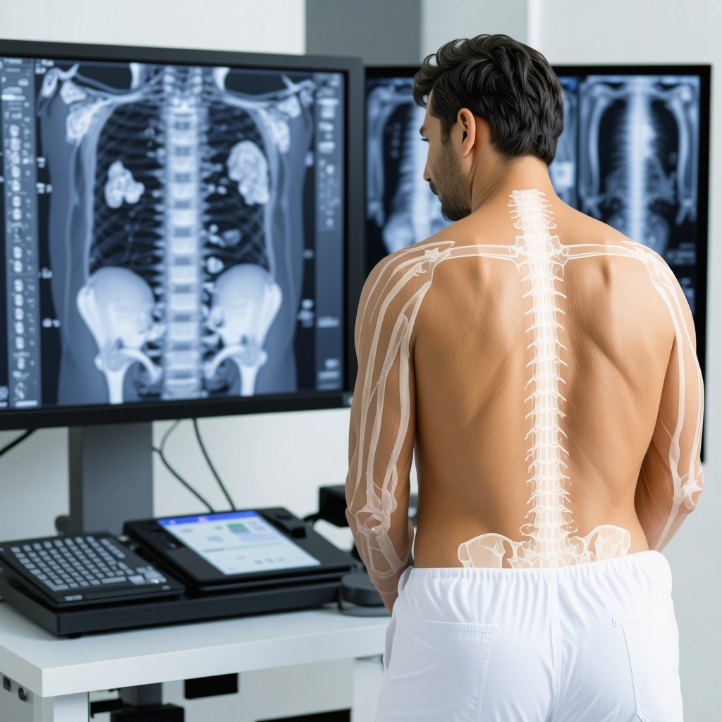
The Diagnostic Dilemma: MRI or X-Ray for Back Pain?
Back pain remains one of the most common complaints driving patients to seek orthopedic care, yet determining the most appropriate imaging modality can be nuanced. While X-rays are often the frontline tool due to accessibility and cost-effectiveness, Magnetic Resonance Imaging (MRI) offers a deeper, more detailed perspective on spinal structures. Understanding when to request an MRI over an X-ray is critical for accurate diagnosis and tailored treatment.
Beyond the Surface: What X-Rays Reveal and What They Miss
X-rays excel at visualizing bone structures, revealing fractures, alignment issues, and advanced degenerative changes. However, they provide limited information about soft tissues such as intervertebral discs, nerve roots, ligaments, and the spinal cord itself. For example, a patient with chronic low back pain and a suspected herniated disc may have an unremarkable X-ray but significant pathology visible on MRI.
What clinical scenarios warrant an MRI instead of or after an X-ray?
MRI is indicated when back pain is accompanied by neurological symptoms like numbness, weakness, or radiating leg pain suggesting nerve root compression. Additionally, MRI is preferred if there is suspicion of infection, tumor, or inflammatory conditions that cannot be detected by X-ray. For instance, a patient presenting with severe, progressive neurological deficits and no obvious bony abnormalities on X-ray should promptly undergo MRI for detailed soft tissue assessment.
Case Study Insight: When MRI Changed the Treatment Path
Consider a middle-aged office worker with persistent lumbar pain initially evaluated with X-rays showing mild degenerative changes. Despite conservative management, symptoms worsened with sciatica development. Subsequent MRI revealed a herniated lumbar disc compressing the sciatic nerve. This imaging insight allowed the orthopedic specialist to tailor a precise non-surgical treatment plan, preventing unnecessary surgery and facilitating targeted physical therapy.
Expert Nuances: Balancing Cost, Accessibility, and Diagnostic Yield
While MRI provides superior soft tissue contrast, it is more expensive and less accessible than X-rays. Therefore, clinical judgment grounded in a thorough history and physical examination guides imaging decisions. In many cases, starting with an X-ray is reasonable, but persistent or complex symptoms should prompt MRI consideration. For a comprehensive discussion on imaging strategies for back pain, see expert recommendations at Back Pain Imaging: MRI vs. X-Ray First Choice Explained.
Integrating Imaging with Holistic Orthopedic Care
Imaging is just one piece of the diagnostic puzzle. Effective management often involves combining imaging findings with clinical expertise, patient history, and other assessments. Patients diagnosed with herniated discs via MRI may benefit from non-surgical orthopedic care options that minimize invasive interventions while maximizing recovery.
For those navigating complex spine issues, choosing the right specialist is essential. Explore guidelines on selecting an orthopedic surgeon for spine care to ensure expert diagnosis and treatment planning.
If you have questions or want to share your experience with back pain diagnostics, feel free to reach out to our expert team or leave a comment below. Your insights could help others make informed decisions about their spine health.
Listening to Your Body: My Experience with Imaging Decisions
Reflecting on my journey with back pain, I remember vividly the uncertainty I felt when my doctor suggested starting with an X-ray. It seemed like a straightforward step, but the pain didn’t improve, and new symptoms started creeping in. That’s when the MRI was recommended, and it felt like a turning point. The detailed images revealed a herniated disc that the X-ray couldn’t capture, guiding a treatment plan tailored exactly to my condition.
The Subtle Signs That Prompted a Deeper Look
What I learned from this experience is how crucial it is to pay attention to subtle symptoms that might indicate the need for more advanced imaging. Tingling in the legs, weakness, or persistent radiating pain are red flags that shouldn’t be ignored. According to a recent article published by the American Academy of Orthopaedic Surgeons, MRI is often recommended when neurological symptoms accompany back pain. This aligns perfectly with my case and many others I’ve spoken to.
How do you know when it’s time to ask for an MRI?
That’s a question many of us grapple with. In my opinion, the answer lies in active communication with your healthcare provider. If your symptoms persist despite initial treatment or if new symptoms like numbness or leg weakness develop, it’s worth discussing the possibility of an MRI. Remember, imaging is a tool to support your care—not the entire story.
Balancing Practicality and Precision in Imaging Choices
Cost and accessibility are real concerns for many, including myself. While MRI offers comprehensive insights, it isn’t always immediately available or covered by insurance. Starting with an X-ray often makes sense, but knowing when to escalate is key. For those interested in a deeper dive, the resource Back Pain Imaging: MRI vs. X-Ray First Choice Explained offers an excellent overview.
Integrating Imaging Results with Holistic Care
What truly made a difference in my recovery was not just the imaging but how my orthopedic team integrated those results with physical therapy and other non-surgical options. If you’re dealing with a herniated disc or similar conditions, exploring effective non-surgical orthopedic care can often provide relief without jumping straight to surgery.
Have you had to decide between MRI and X-ray for your back pain? What guided your choice? I’d love to hear your story or any questions you might have. Share your experiences in the comments below or reach out through our contact page. Together, we can navigate these decisions with greater confidence.
Decoding Complex Back Pain: Advanced MRI Techniques Beyond Conventional Imaging
While traditional MRI protocols provide exceptional visualization of spinal anatomy, recent advancements in imaging techniques have elevated diagnostic precision for complex back pain cases. Techniques such as diffusion tensor imaging (DTI) and functional MRI (fMRI) enable clinicians to assess nerve integrity and muscle activation patterns that conventional MRI might miss. These modalities can reveal microstructural changes in nerve roots and subtle inflammatory processes, often correlating with refractory symptoms despite normal standard imaging.
Incorporating these advanced MRI sequences into diagnostic algorithms can be particularly transformative for patients with persistent radiculopathy or ambiguous presentations where standard imaging fails to elucidate the source of pain. This nuanced approach demands both sophisticated equipment and specialized interpretation skills, underscoring the importance of referral to centers with expertise in spine imaging.
How can diffusion tensor imaging improve detection of nerve root pathology in chronic back pain?
Diffusion tensor imaging (DTI) measures the directional movement of water molecules along nerve fibers, allowing visualization of nerve tract integrity. In chronic back pain patients with suspected nerve root compression, DTI can detect microstructural damage or demyelination not apparent on standard MRI. This capability enhances diagnostic confidence in distinguishing between mechanical compression and neuropathic pain origins, guiding more targeted interventions. Recent studies published in NeuroImage: Clinical illustrate how DTI metrics correlate with symptom severity and postoperative outcomes, making it a promising adjunct for complex cases.
Integrating Imaging Findings with Electrophysiological Studies for Comprehensive Assessment
High-resolution imaging alone cannot always capture the functional status of spinal nerves. Electromyography (EMG) and nerve conduction studies (NCS) remain invaluable complements, especially when MRI findings are equivocal. When combined, imaging reveals anatomical abnormalities while electrophysiological tests assess nerve function, enabling a holistic assessment of the neuro-musculoskeletal interface.
This integrated diagnostic paradigm is crucial when planning surgical interventions or evaluating candidates for advanced therapies such as spinal cord stimulation. Orthopedic specialists increasingly advocate for a multidisciplinary approach, incorporating radiologists, neurologists, and physical therapists to interpret the full clinical picture.
Economic and Ethical Considerations in Advanced Imaging Utilization
While the clinical benefits of advanced MRI techniques are compelling, the cost-effectiveness and accessibility challenges cannot be overlooked. Insurance coverage for specialized imaging varies widely, and the risk of incidental findings can lead to patient anxiety and unnecessary procedures. Ethical stewardship mandates judicious use of these resources, balancing diagnostic yield against potential harms.
Shared decision-making, informed by detailed patient education about the benefits and limitations of advanced imaging, is essential. Clinicians must remain vigilant against overutilization, ensuring imaging requests are aligned with validated clinical indications and not driven by defensive medicine or patient pressure alone.
Next Steps: Harnessing Artificial Intelligence to Enhance Imaging Interpretation
The future of back pain imaging lies in the integration of artificial intelligence (AI) tools that augment radiologist expertise. Machine learning algorithms are being developed to automatically detect subtle disc herniations, spinal stenosis, and nerve root impingements with high accuracy and reproducibility. These technologies promise to reduce diagnostic delays and inter-observer variability, especially in community settings with limited subspecialty radiology support.
Early clinical trials demonstrate AI-enhanced MRI interpretations can stratify patients by risk and predict responsiveness to conservative versus surgical treatment, optimizing individualized care pathways. As these tools mature, orthopedic practitioners will need to engage actively with AI literacy to harness their full potential responsibly.
If you’re navigating complex back pain diagnostics or interested in how cutting-edge imaging can refine your care, connect with our expert team to explore tailored diagnostic strategies that integrate the latest advances.
Unlocking Next-Level Insights: Advanced MRI Modalities in Spine Diagnostics
Emerging MRI technologies are redefining the diagnostic landscape for patients with persistent or complex back pain. Beyond conventional imaging, advanced techniques such as diffusion tensor imaging (DTI) and functional MRI (fMRI) provide unparalleled visualization of neural microstructure and functional dynamics within the spine. These modalities enable clinicians to detect subtle pathologies like nerve fiber disruption or inflammatory processes that standard MRI sequences often overlook.
How can diffusion tensor imaging improve detection of nerve root pathology in chronic back pain?
DTI capitalizes on the anisotropic diffusion of water molecules along nerve fibers, quantitatively mapping nerve integrity. In chronic back pain with suspected radiculopathy, DTI facilitates identification of demyelination or microstructural abnormalities not visible on routine imaging. This enhances differential diagnosis between mechanical nerve compression and neuropathic pain syndromes, informing precision treatment plans. Peer-reviewed research in NeuroImage: Clinical demonstrates significant correlations between DTI metrics and clinical symptomatology, underscoring its clinical utility.
Synergizing Imaging with Electrophysiological Assessments for Holistic Evaluation
While advanced imaging elucidates anatomical anomalies, integrating electrophysiological tests such as electromyography (EMG) and nerve conduction studies (NCS) offers a comprehensive appraisal of nerve function. This multifaceted approach bridges the gap between structural findings and functional impairment, crucial for nuanced clinical decision-making. Such integration is particularly valuable in ambiguous cases where imaging alone cannot conclusively characterize nerve involvement.
Ethical and Economic Dimensions of Deploying Cutting-Edge Imaging Tools
Despite the diagnostic advantages, advanced MRI modalities pose challenges related to cost, accessibility, and incidental findings that may precipitate patient anxiety or unwarranted interventions. Ethical stewardship mandates that clinicians employ these technologies judiciously, guided by robust clinical indications and shared decision-making frameworks. Transparency about potential benefits and limitations ensures patient-centered care and resource optimization.
Harnessing Artificial Intelligence: The Future of Spine Imaging Interpretation
Artificial intelligence (AI) is rapidly advancing the field of spinal imaging by augmenting diagnostic accuracy and efficiency. Machine learning algorithms trained on large datasets can automate detection of subtle disc herniations, spinal stenosis, and nerve root impingements with remarkable precision. This technological synergy reduces inter-observer variability and expedites diagnostic workflows, particularly in settings with limited specialist availability.
Early clinical validations suggest AI-enhanced MRI analyses can stratify patients based on prognosis and predict therapeutic responsiveness, thereby personalizing treatment strategies. Orthopedic practitioners are encouraged to engage with AI tools proactively, ensuring ethical integration and maximizing clinical benefit.
If you are navigating complex back pain or interested in how these advanced imaging innovations can refine your diagnostic and treatment approach, connect with our expert team to access cutting-edge, individualized care pathways.

Frequently Asked Questions (FAQ)
1. When should I ask my doctor for an MRI instead of an X-ray for back pain?
If your back pain is accompanied by neurological symptoms such as numbness, weakness, or radiating leg pain, or if initial conservative treatments fail, it is advisable to discuss an MRI with your doctor. MRI provides detailed images of soft tissues and nerve structures that X-rays cannot visualize.
2. Can X-rays detect herniated discs or nerve compression?
No, X-rays primarily show bone structures and cannot directly visualize soft tissues like intervertebral discs or nerve roots. MRI is the preferred modality to assess these structures and detect conditions such as herniated discs or nerve impingement.
3. What are diffusion tensor imaging (DTI) and functional MRI (fMRI), and how do they improve back pain diagnosis?
DTI and fMRI are advanced MRI techniques that provide insights into nerve fiber integrity and functional muscle activation, respectively. They can detect microstructural nerve damage and subtle inflammatory changes not seen on conventional MRI, aiding in more precise diagnosis of complex or refractory back pain.
4. How do electrophysiological studies complement MRI findings?
Electromyography (EMG) and nerve conduction studies (NCS) assess nerve function and can detect abnormalities even when MRI findings are inconclusive. When combined, these tests offer a comprehensive evaluation of the anatomical and functional status of spinal nerves.
5. Are advanced MRI techniques widely available and covered by insurance?
Availability and insurance coverage for advanced MRI modalities vary by region and provider. These techniques often require specialized equipment and expertise, and may not be routinely covered. Discuss the benefits and costs with your healthcare provider to make an informed decision.
6. What ethical considerations exist regarding the use of advanced imaging?
Advanced imaging should be used judiciously to avoid unnecessary costs, patient anxiety from incidental findings, and unwarranted interventions. Shared decision-making and adherence to clinical guidelines help ensure imaging is appropriate and patient-centered.
7. How is artificial intelligence (AI) transforming back pain imaging?
AI algorithms enhance MRI interpretation by detecting subtle spinal abnormalities with high accuracy and consistency. This technology reduces diagnostic delays and variability, helping tailor personalized treatment plans, especially in settings lacking specialized radiologists.
8. Can imaging alone determine the best treatment for back pain?
No, imaging findings are only one part of the diagnostic process. Clinical examination, patient history, and other tests must be integrated to design effective, individualized treatment strategies.
9. What should patients do if their symptoms persist despite normal X-rays?
Persistent or worsening symptoms warrant further evaluation, often with MRI and possibly electrophysiological studies, to uncover soft tissue or nerve-related causes that X-rays cannot detect.
10. How can patients ensure they receive appropriate imaging and care?
Active communication with healthcare providers, understanding symptom red flags, and seeking care from specialists experienced in spine diagnostics can help patients receive timely and accurate imaging and treatment.
Trusted External Sources
- American Academy of Orthopaedic Surgeons (AAOS) – OrthoInfo: Provides evidence-based guidelines on diagnosing and managing back pain, including imaging recommendations that balance clinical indications and resource use.
- NeuroImage: Clinical (Peer-Reviewed Journal): Publishes advanced neuroimaging research such as diffusion tensor imaging studies that deepen understanding of nerve pathology in back pain.
- National Institute of Neurological Disorders and Stroke (NINDS): Offers comprehensive information on spinal disorders, diagnostic techniques, and emerging imaging technologies relevant to back pain.
- Radiological Society of North America (RSNA): Delivers expert resources on imaging modalities, technical advances, and best practices in spinal imaging.
- Centers for Medicare & Medicaid Services (CMS): Provides insights into coverage policies and economic considerations surrounding advanced imaging procedures.
Conclusion
Choosing between MRI and X-ray for back pain diagnosis requires a nuanced approach that integrates symptomatology, clinical findings, and patient-specific factors. While X-rays remain valuable for initial bone assessment, MRI’s superior soft tissue visualization is indispensable when neurological involvement or complex pathology is suspected. Advanced MRI modalities such as diffusion tensor imaging and functional MRI, alongside electrophysiological studies, elevate diagnostic precision for challenging cases. Ethical and economic considerations underscore the necessity of judicious imaging use, supported by shared decision-making. The integration of artificial intelligence promises to further enhance diagnostic accuracy and personalized care. Ultimately, a holistic, expert-guided strategy that couples imaging insights with clinical expertise optimizes outcomes for those suffering from back pain. Engage with your healthcare providers, explore these advanced diagnostic options, and share your experiences to contribute to a well-informed community dedicated to spine health.
This post really highlights the complexity of deciding between MRI and X-ray when dealing with back pain. In my own experience, I initially had an X-ray for persistent lower back discomfort, which showed some mild degenerative changes but nothing alarming. It wasn’t until I developed tingling in my leg that my doctor recommended an MRI, which revealed a herniated disc pressing on a nerve root — something an X-ray just couldn’t show. What struck me was how crucial it is to listen to those subtle neurological symptoms that might warrant deeper imaging. However, I also appreciate the point about balancing the cost and accessibility of MRIs versus the more readily available X-rays. Has anyone else found that communication with their healthcare provider about new or worsening symptoms was the key to getting the appropriate imaging done? How do you navigate this balance between being proactive about your health and the practical constraints like insurance and wait times? I’d love to hear how others have managed this decision-making process in their own back pain journeys.
Marilyn, your experience resonates deeply with me, especially regarding the challenge of navigating communication with healthcare providers amidst symptoms that evolve or persist. In my case, my initial back pain was dismissed with just an X-ray after a minor strain was suspected. It wasn’t until persistent numbness and occasional weakness in my foot emerged that I advocated for an MRI, which revealed nerve root compression caused by a bulging disc invisible on the X-ray. What made the difference was preparing thorough notes about symptom progression and the impact on daily life before appointments. Sharing detailed experiences helped my physician appreciate the urgency for advanced imaging despite initial insurance hurdles and wait times.
I also see this post’s point about how clinical judgment—balancing cost, accessibility, and diagnostic precision—is crucial. It’s a fine line between being proactive and respecting system limitations. Some providers are more receptive to patient input, while others rely heavily on strict imaging protocols. How do others here prepare or advocate effectively during consultations to ensure their symptoms’ nuances are acknowledged? Additionally, has anyone found resources or strategies that help navigate insurance authorization for MRIs when symptoms are subtle but persisting? Ultimately, fostering a collaborative dialogue seems key to timely, tailored imaging and treatment.
This post offers a thorough overview of the nuanced decision-making involved in back pain diagnostics. I find that my own experience aligns with the emphasis on knowing when to escalate to MRI—initially, I was advised to start with X-rays, which showed mild degenerative changes. It was only after persistent symptoms, including numbness and radiating pain, that my doctor recommended an MRI, revealing a herniated disc pressing on a nerve. Paying attention to subtle neurological signs proved crucial in guiding the right imaging choice. It raises an interesting point about patient advocacy and active communication with healthcare providers, especially when symptoms evolve or worsen. Have others found that maintaining detailed symptom records or preparing questions before appointments helps in expediting appropriate imaging? Also, given the advances in MRI technology like DTI and functional MRI discussed here, I wonder how accessible these techniques are across different healthcare settings and whether they are becoming standard practice for complex cases. Would love to hear experiences from those who’ve navigated similar challenges—sharing insights can really empower others dealing with inconclusive initial assessments.
Reading through the detailed explanations about when to choose MRI over X-rays for back pain really resonated with me. I had a somewhat tricky case where my initial X-rays were inconclusive despite ongoing lower back pain and some numbness in my leg. Like the case study described, it was only after an MRI that a herniated disc pressing on a nerve root was diagnosed, which explained my symptoms better than the X-ray ever could. What stands out to me is the delicate balance between cost and accessibility versus diagnostic precision. It’s easy to feel caught between wanting the most accurate diagnosis and grappling with insurance and wait times. I’ve found that preparing a clear timeline of symptom progression and emphasizing any neurological changes during appointments helps providers appreciate the need for an MRI sooner rather than later. I’m curious, for others who have navigated this, how have you managed to advocate effectively within healthcare constraints? Also, has anyone had experiences with advanced imaging techniques like diffusion tensor imaging and felt it made a measurable difference in understanding complex back issues? It seems promising but might still be hard to access. Overall, this post underscores how vital open communication and informed decision-making are in managing back pain diagnostics.
This article really emphasizes how critical it is to consider the full clinical picture when deciding between MRI and X-ray for back pain. In my experience, I initially underwent X-rays which showed only mild degenerative changes, but my symptoms worsened and included radiating pain and numbness. It was only after an MRI that a herniated disc was diagnosed, which the X-ray couldn’t reveal. This highlights how soft tissue injuries can be missed without advanced imaging. I wonder, with the growing availability of point-of-care ultrasound, could that be a helpful intermediate step in evaluating soft tissue concerns before resorting to MRI? Also, how do others manage situations where MRI isn’t immediately accessible due to wait times or costs? Is there an effective way to advocate for necessary imaging based on evolving symptoms? Sharing strategies on navigating these challenges could really help others in similar situations. Ultimately, listening to subtle signs and advocating for comprehensive assessment seem vital for proper diagnosis and treatment.