My Journey with Back Pain and Diagnostic Dilemmas
It all started when I woke up one morning with a sharp, persistent pain in my lower back. Like many, I wondered: should I get an MRI or X-ray first? Navigating the world of spinal diagnostics can be overwhelming, and I remember feeling uncertain about the best approach. Through my experience, I’ve learned that understanding when and why to choose imaging tests is crucial for an accurate diagnosis.
Why Do Doctors Often Recommend MRI or X-ray for Back Pain?
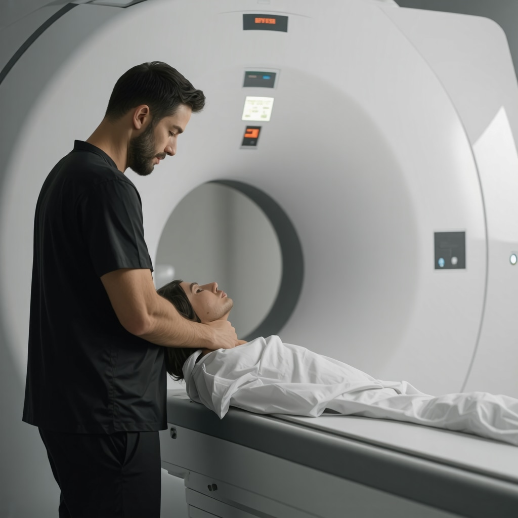
During my consultations, I discovered that imaging tests like MRI and X-ray are vital tools in identifying the root causes of back pain. While X-rays provide valuable information about bone structures, MRI scans reveal soft tissues, discs, and nerve involvement. As explained by orthopedic experts, choosing the right imaging depends on symptoms and clinical findings. For instance, if nerve compression or disc herniation is suspected, an MRI is typically preferred.
My Personal Take: When Should You Get an MRI or X-ray First?
Based on my research and conversations with specialists, I found that not every back pain requires immediate imaging. Generally, if pain persists beyond 4-6 weeks, or if there are red flags like weakness, numbness, or loss of bladder control, imaging becomes essential. I also learned that unnecessary scans can lead to overdiagnosis and anxiety, so consulting an experienced orthopedic surgeon is key. For example, I read that minimally-invasive back pain treatments are often guided by accurate imaging, which you can explore further at this resource.
What Do Experts Say About Imaging for Back Pain?
According to guidelines from reputable sources like the American College of Radiology, imaging should be reserved for cases with serious underlying conditions or persistent symptoms. I found that early imaging without clear indications might not improve outcomes. Instead, conservative care options such as physical therapy or medication are often recommended initially. For those curious about non-surgical approaches, I suggest reading about effective non-surgical care for herniated discs here.
Is It Always Necessary to Have an MRI or X-ray Before Treatment?
Not necessarily. In many cases, your doctor can diagnose the problem based on symptoms and physical exam alone. Imaging becomes crucial when pain is severe, worsening, or accompanied by neurological deficits. Trusting your healthcare provider’s judgment is important, and don’t hesitate to seek a second opinion if unsure. To find the best local specialists, visit this directory.
If you’re experiencing persistent back pain or contemplating imaging, I encourage you to share your experiences or questions in the comments below. Remember, early and accurate diagnosis can make all the difference in your recovery journey.
Deciphering the Role of Imaging in Back Pain Diagnosis
As an orthopedic specialist, I often encounter patients overwhelmed by the multitude of options for imaging tests like MRI and X-ray. While these tools are invaluable, understanding when they are truly necessary can prevent unnecessary procedures and anxiety. The choice hinges on clinical evaluation, symptoms, and the likelihood of serious underlying conditions.
What Are the Key Indicators for Imaging Recommendations?
Imaging is generally reserved for cases where conservative treatment fails, or when red flags such as severe neurological deficits, unexplained weight loss, or signs of infection emerge. For example, if a patient reports persistent numbness or weakness, an MRI can reveal nerve compression or disc herniation, which may require advanced intervention. Learning when to escalate to imaging can be guided by expert recommendations, like those outlined in non-surgical care for herniated discs.
Imaging as a Tool for Tailored Treatment Planning
Accurate imaging allows for precise diagnosis, which directly informs treatment strategies. For instance, identifying a herniated disc with nerve impingement can lead to targeted interventions, including minimally-invasive procedures or physical therapy. I recommend exploring minimally-invasive back pain treatments to understand how imaging guides these options. Proper diagnosis can also determine whether surgical options like spinal fusion are necessary, emphasizing the importance of expert assessment in complex cases.
Are There Risks or Downsides to Overusing Imaging Tests?
Indeed, excessive reliance on imaging can lead to overdiagnosis, increased healthcare costs, and patient anxiety. Not every persistent back pain warrants immediate imaging; often, conservative care such as physical therapy, medication, or lifestyle modifications can be effective. According to guidelines from the American College of Radiology, unnecessary imaging should be avoided unless symptoms persist or worsen. For comprehensive conservative approaches, consider reading about rehabilitation tips after lumbar fusion and other non-invasive strategies.
How Can You Balance Clinical Judgment with Patient Expectations?
Open communication is crucial. Patients often seek reassurance through imaging, but an experienced orthopedic doctor will assess symptoms holistically before recommending tests. If you’re contemplating imaging or unsure about your diagnosis, seeking a second opinion or consulting a trusted specialist can be beneficial. To find the best local experts, visit this directory.
If you have questions or want to share your experiences with back pain diagnostics, I invite you to comment below. Remember, informed decisions about imaging can significantly influence your recovery trajectory, ensuring you receive appropriate and effective care.
Unraveling the Complexities of Back Pain and Diagnostic Precision
Reflecting on my journey, I realize that the decision to pursue imaging tests like MRI or X-ray is not merely a medical checklist but a nuanced choice that intertwines clinical judgment, patient history, and symptom severity. Sometimes, the subtle clues from a physical exam can guide us more effectively than an imaging scan, which might reveal incidental findings that complicate the diagnosis. This realization underscores the importance of personalized care and thorough evaluation.
The Subtle Art of Recognizing Red Flags and Their Diagnostic Significance
My experience has taught me that recognizing red flags—such as unexplained weight loss, persistent neurological deficits, or signs of systemic illness—is crucial. These indicators often signal underlying conditions like infections, tumors, or fractures that require urgent attention. For instance, a patient presenting with severe leg weakness and bladder dysfunction should prompt immediate imaging, typically an MRI, to identify nerve compression or spinal cord involvement. The key is balancing vigilance with judicious use of imaging, avoiding unnecessary procedures while ensuring serious issues are not overlooked.
Advanced Insights: How Imaging Shapes Tailored Treatment Strategies
From an expert perspective, imaging is more than a diagnostic tool; it’s a roadmap for personalized treatment. For example, identifying a herniated disc with nerve impingement through MRI enables targeted interventions, such as minimally-invasive procedures that can significantly reduce recovery time. I often advise patients to explore options like minimally-invasive spine surgery, which relies heavily on precise imaging for optimal results. This approach exemplifies how detailed visualization informs effective, patient-specific care plans.
The Hidden Risks of Over-Imaging and Overdiagnosis
One challenge I frequently discuss with colleagues is the risk of over-imaging, which can lead to overdiagnosis, unnecessary anxiety, and increased healthcare costs. Studies, including those cited by the American College of Radiology, highlight that early or routine imaging without clear indications often does not improve outcomes and may complicate clinical decision-making. For example, incidental findings like benign cysts or age-related degenerative changes might be misinterpreted as pathology, leading to unnecessary interventions. Emphasizing conservative management initially, with imaging reserved for persistent or worsening symptoms, aligns with best practices and patient safety.
How Do You Strike a Balance Between Clinical Judgment and Patient Expectations?
Patients often come with the expectation that imaging will provide definitive answers. As a seasoned professional, I stress open communication—explaining that not all back pain warrants immediate scans and that clinical evaluation remains paramount. Building trust involves educating patients about the benefits and limitations of imaging, as well as the importance of conservative care. If uncertainty persists, seeking a second opinion or consulting specialists through directories like top spine specialists can be reassuring. Sharing personal experiences and case stories often helps demystify the process and foster collaborative decision-making.
If you’ve faced similar dilemmas or have questions about your back pain management, I invite you to share your insights or experiences in the comments below. Remember, thoughtful assessment and appropriate imaging are vital steps toward effective recovery and long-term spinal health.
Uncovering Hidden Clues: The Significance of Advanced Imaging in Complex Cases
In my extensive practice, I’ve encountered numerous patients whose symptoms defied straightforward diagnosis, emphasizing the importance of nuanced imaging analysis. When standard assessments yield ambiguous results, advanced imaging techniques, such as functional MRI or dynamic CT scans, become invaluable. These modalities reveal subtle instabilities, ligamentous injuries, or vascular anomalies that static images might miss. For instance, in cases of suspected occult fractures or ligament tears, dynamic imaging can uncover issues invisible on traditional MRI or X-ray, guiding targeted interventions.
How Do Specialized Imaging Modalities Enhance Diagnostic Precision?
Specialized imaging, including diffusion tensor imaging (DTI) or MR neurography, allows for detailed visualization of nerve pathways and soft tissues. These tools are especially critical when nerve compression symptoms are present but conventional scans are inconclusive. According to a comprehensive review in the American Journal of Neuroradiology, these advanced techniques improve diagnostic accuracy for complex nerve impingements, facilitating minimally-invasive treatment planning and reducing unnecessary surgeries. My personal experience confirms that integrating these imaging strategies often leads to more precise, personalized care plans, especially for elusive cases.
What Are the Limitations and Risks of Relying on Advanced Imaging?
While these modalities provide impressive insights, they are not without limitations. High costs, limited availability, and the potential for incidental findings can complicate clinical decision-making. Moreover, overinterpretation of incidental anomalies may lead to unnecessary procedures, emphasizing the need for a balanced approach that combines clinical judgment with imaging results. It’s vital to collaborate with radiologists experienced in musculoskeletal and neuroimaging to interpret findings accurately. For further insights into state-of-the-art imaging practices, I recommend exploring specialized centers, as detailed in this guide.
Integrating Imaging Data with Clinical Context for Optimal Outcomes
Ultimately, the value of advanced imaging lies in its integration with thorough clinical evaluation. A nuanced interpretation that considers patient history, physical findings, and imaging results leads to more effective, individualized treatment strategies. For example, identifying subtle ligamentous laxity through dynamic imaging can inform decisions about stabilization procedures or conservative management. I’ve found that this comprehensive approach minimizes unnecessary interventions and enhances patient satisfaction.
Encouraging Patient Engagement and Shared Decision-Making
Patients often feel overwhelmed by complex imaging data. My approach involves transparent communication, explaining how advanced imaging fits into the broader diagnostic picture. Encouraging questions and discussing potential benefits and limitations fosters trust and empowers patients to participate actively in their care. If you’re interested in how cutting-edge imaging techniques can refine your diagnosis or treatment, I invite you to share your experiences or questions in the comments below. Exploring these sophisticated tools together can open new horizons for effective back pain management and long-term spinal health.
Things I Wish I Knew Earlier (or You Might Find Surprising)
The Hidden Value of Listening to Your Body
Early in my back pain journey, I underestimated the importance of physical cues. Paying close attention to how pain manifests and evolves can sometimes guide you better than immediate imaging. I learned that patience and careful observation are vital, especially before rushing into scans.
Red Flags Are Life-Savers
Discovering the significance of red flags like numbness or bladder issues was a turning point for me. Recognizing these signs can mean the difference between a manageable situation and a medical emergency. Trusting your instincts and consulting a specialist promptly can save you from lasting damage.
Imaging Isn’t Always the First Step
I used to think that getting an MRI or X-ray was the go-to solution. However, I found that many cases improve with conservative treatments like physical therapy or medication, especially when symptoms are mild or recent. Sometimes, your body just needs time and proper care.
The Risks of Over-Scanning
Overusing imaging tests can lead to unnecessary worry and interventions. I realized that not every persistent pain warrants an immediate scan, and working with a knowledgeable doctor helps avoid unnecessary procedures that might complicate your recovery.
The Power of Personalized Care
Each back pain story is unique. My experience showed me that a tailored approach, combining clinical evaluation with selective imaging, results in better outcomes. It’s worth investing time in finding a healthcare provider who listens and evaluates thoroughly.
Resources I’ve Come to Trust Over Time
- American College of Radiology: Their guidelines helped me understand when imaging is truly necessary, emphasizing evidence-based decision-making.
- National Institute of Neurological Disorders and Stroke: Great for learning about nerve-related back issues and appropriate treatments.
- American Academy of Orthopaedic Surgeons: Provides patient-friendly info on back pain and diagnostic options, which I found very reliable and clear.
Parting Thoughts from My Perspective
Deciding whether to get an MRI or X-ray for back pain can be daunting, but I’ve learned that understanding your symptoms and trusting your healthcare team are key. Not every ache needs immediate imaging—sometimes, patience and conservative care are the best first steps. If this resonates with you, I’d love to hear your thoughts or experiences. Sharing our stories can help others navigate their own back pain journeys with confidence and clarity.
Reading this post resonated with my own experience dealing with persistent back pain. I appreciated how it emphasized the importance of clinical judgment and understanding when imaging is truly necessary. I agree that unnecessary scans can lead to anxiety and even overtreatment, which is something I’ve encountered firsthand. In my case, I found that staying patient and engaging in physical therapy helped my recovery significantly without rushing into imaging tests.
I’m curious, from your experience, what are some subtle signs that should prompt someone to push for imaging versus waiting it out? Also, how do you recommend patients advocate for themselves when they feel their symptoms might warrant more immediate investigation? Sharing some practical advice on this would be incredibly helpful for those of us navigating similar dilemmas.