My Journey into Orthopedic Diagnostic Imaging: Why Preparation Matters
Last year, I found myself nervously awaiting the results of an orthopedic scan. Having experienced persistent back pain, I knew that accurate imaging was crucial for proper diagnosis and treatment. My journey into understanding how to prepare for orthopedic diagnostic imaging taught me valuable lessons I want to share with anyone facing similar situations.
Understanding the Importance of Proper Preparation
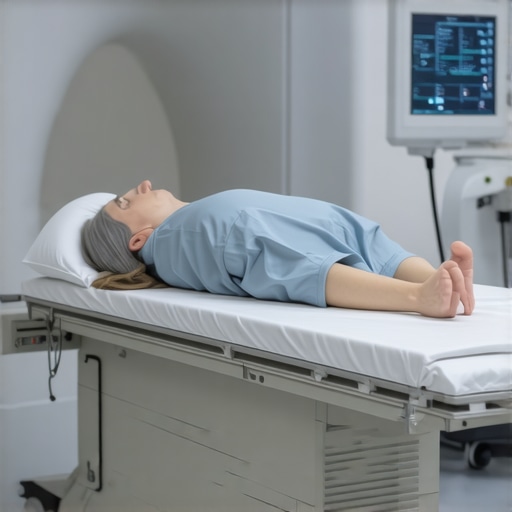
Preparing for your scan isn’t just about showing up on time; it’s about ensuring that the images captured are clear and precise. I learned that factors like fasting, clothing choices, and informing your technician about any implants or recent injuries can significantly influence the accuracy of your results. For instance, wearing loose, comfortable clothing and avoiding metal accessories helped facilitate a smoother imaging process.
What Should You Know Before Your Scan?
How can I ensure I’m fully prepared for my orthopedic imaging?
From my experience, the best way to prepare is to follow your doctor’s instructions carefully. They might recommend specific fasting protocols or advise on medication adjustments. Reading up on the procedure beforehand can also ease anxiety—knowing what to expect makes the process less intimidating. I also found that arriving early and bringing any relevant medical history or previous imaging reports can help your healthcare team provide the best possible care.
For additional insights, I recommend checking out resources like the American College of Radiology’s guidelines on imaging preparation. They emphasize that proper preparation not only improves image quality but also reduces the need for repeat scans, saving you time and exposure to unnecessary radiation.
Gaining Accurate Results: My Personal Tips
During my scan, I was surprised at how much of a difference proper positioning made. The technician took the time to help me stay still and in the correct position, which is vital for detailed images. I also appreciated the importance of honest communication—if you’re uncomfortable or have concerns, speaking up ensures your safety and the quality of the scan.
Post-scan, I learned that understanding the results can be daunting. However, discussing the images with your orthopedic specialist and asking questions about findings and next steps can make a significant difference. If you’re unsure or need a second opinion, don’t hesitate to explore options like a second opinion from a trusted orthopedic surgeon here.
Why Accurate Imaging is a Cornerstone of Effective Orthopedic Care
Accurate diagnostic imaging is foundational for effective treatment planning. It allows your doctor to pinpoint issues precisely, whether it’s a herniated disc, ligament tear, or bone fracture. I’ve realized that investing time in preparation pays off by providing clearer images and, ultimately, better care.
Are you curious about how advancements in imaging technology are shaping the future of orthopedic diagnostics? Technologies like 3D imaging and MRI improvements are making diagnostics more precise than ever, according to recent reports from trusted sources like RadiologyInfo.org.
Feeling empowered after my experience, I encourage others to approach their imaging appointments with confidence and preparation. If you’re interested in exploring more about orthopedic imaging or need guidance, I’m here to share and learn together. Drop a comment below or share your experiences—your story might help someone else navigate their journey more smoothly.
Unlocking the Secrets to Clearer Orthopedic Imaging: An Expert’s Perspective
When it comes to diagnosing complex spinal and joint issues, the quality of your imaging can make all the difference. As an orthopedic specialist, I’ve seen firsthand how proper preparation can significantly enhance the clarity and accuracy of diagnostic scans, leading to better treatment decisions. Whether you’re preparing for an MRI, CT scan, or X-ray, understanding the nuances of imaging prep is crucial for optimal results.
The Nuances of Pre-Scan Preparation: What Every Patient Should Know
Preparation goes beyond simply arriving at the imaging center on time. It involves a series of steps tailored to your specific procedure. For example, fasting might be required before an MRI to reduce artifacts caused by digestive activity, while removing metallic objects is essential to prevent image distortion. Wearing loose, comfortable clothing without metal accessories also aids in swift scanning and high-quality images. These details, often overlooked, are vital for radiologists and orthopedic surgeons to interpret your images accurately.
Expert Strategies for Ensuring Your Imaging Is Flawless
From my clinical experience, communicating openly with your imaging technician is invaluable. If you have implants, pacemakers, or other medical devices, informing the staff beforehand allows them to adjust protocols accordingly. Additionally, arriving early provides time to review your medical history and ensure all relevant documents are available. This proactive approach minimizes the chances of needing repeat scans, which can delay diagnosis and increase exposure to radiation in some cases.
For a comprehensive understanding of how to prepare, I recommend reviewing resources like the American College of Radiology’s guidelines, which emphasize the importance of tailored preparation for different imaging modalities. Proper prep not only improves image quality but also reduces patient anxiety and streamlines the entire process, making your experience more comfortable and efficient.
Decoding the Images: How Preparation Impacts Diagnostic Precision
Accurate interpretation hinges on high-quality images. For instance, a well-positioned MRI can reveal subtle disc herniations or ligament tears that might otherwise be missed. During my practice, I’ve observed that patients who follow pre-scan instructions tend to receive more precise diagnoses, enabling targeted treatments—be it conservative management or surgical intervention.
Have you ever wondered how technological innovations are elevating orthopedic diagnostics? Advances such as 3D imaging and high-resolution MRI are transforming the landscape, making it possible to detect issues earlier and with greater confidence. According to recent reports from RadiologyInfo.org, these developments are setting new standards for accuracy and patient outcomes.
What Can You Do to Prepare Better Than the Rest?
Beyond following standard instructions, consider maintaining a detailed medical record of your previous imaging and treatments. Sharing this with your healthcare team can prevent unnecessary repeat scans and ensure they interpret your images within the full context of your medical history. To streamline your journey, explore options like a second opinion from a trusted orthopedic surgeon here.
Interested in learning more about non-invasive treatments that complement clear diagnostics? Visit this resource for insights on rehab strategies following imaging-confirmed diagnoses.
Join the Conversation: Share Your Imaging Experiences
Have you experienced a situation where proper preparation led to a faster diagnosis or better treatment? Or perhaps you faced challenges due to inadequate prep? Sharing your story can help others navigate their own orthopedic journeys more confidently. Feel free to comment below or suggest more topics you’d like to explore—your input might just make a difference in someone else’s health journey.
Deepening the Understanding: The Art and Science of Imaging Preparation
Reflecting on my journey, I realize that preparation for orthopedic diagnostic imaging is not just about following instructions but about understanding the intricate details that influence the quality of your scans. For instance, did you know that even the slightest movement during an MRI can blur the image, potentially leading to misdiagnosis? This is why immobilization techniques and patient comfort are vital topics that often go overlooked but can dramatically affect your results.
Unveiling the Hidden Factors Behind Image Clarity
Through my experience, I’ve discovered that factors like hydration levels, recent physical activity, and even emotional stress can subtly impact imaging outcomes. Dehydration, for example, can cause tissues to appear less distinct, while anxiety can lead to involuntary movements. Being mindful of these nuances, I started adopting calming routines before my scans—deep breathing exercises and staying well-hydrated—which surprisingly improved my experience and the clarity of the images.
The Nuances of Advanced Imaging Technologies and Patient Role
Advancements such as functional MRI (fMRI) and high-resolution 3D imaging are pushing the boundaries of diagnostic precision. As a patient, understanding how these technologies work and how your preparation supports their effectiveness can empower you. For example, knowing that certain sequences require specific positioning or that contrast agents necessitate particular pre-scan protocols made me more proactive in my preparation. This knowledge helps bridge the gap between technological potential and patient participation, ultimately leading to more accurate diagnoses.
What are the lesser-known yet impactful preparation tips that could elevate your imaging results?
One often overlooked tip is the importance of detailed medical documentation. Sharing previous imaging, surgical history, or even specific injury details can guide radiologists in refining their technique. Additionally, discussing any concerns about claustrophobia or discomfort with your technician beforehand allows them to tailor the process—perhaps by offering a mild sedative or adjusting the environment—which can significantly enhance your cooperation and the resulting image quality. For comprehensive guidance, I recommend reviewing this resource.
From Good to Great: Personal Strategies for Optimal Imaging Outcomes
In my own practice, I found that arriving early and mentally preparing myself reduces stress, which directly correlates with better image quality. I also made it a point to communicate openly with the technologists—sharing any recent injuries, implants, or medications that might affect imaging. This proactive approach not only minimizes repeat scans but also ensures your safety and comfort throughout the procedure. Remember, your active role in preparation can make a tangible difference in your healthcare journey.
Technology and Human Factors: A Symbiotic Relationship
As I continue exploring the future of orthopedic diagnostics, I’m excited by how innovations like AI-enhanced image analysis promise even greater accuracy. However, these advancements still rely on high-quality data, which begins with proper patient preparation. My advice? Embrace a mindset of partnership with your healthcare team—understanding that your cooperation, informed by a little knowledge and mindfulness, can significantly influence your diagnostic success.
For those interested in learning more about how to optimize their preparation, visiting this page offers valuable insights on navigating the complexities of orthopedic care and ensuring your imaging helps pave the way for effective treatment.
Join the Conversation: Share Your Techniques and Experiences
Have you discovered any personal tips that improved your imaging results? Or faced unexpected challenges that taught you something new? Sharing your experiences can empower others to approach their scans with confidence and knowledge. Feel free to comment below or explore more on the importance of preparation in your orthopedic journey—your story might inspire someone else to achieve better health outcomes.
Refining Your Approach to Orthopedic Diagnostic Imaging: Insights from a Specialist’s Perspective
Embarking on my journey through the intricacies of orthopedic diagnostic imaging, I quickly realized that meticulous preparation is pivotal for obtaining images that truly reflect the underlying pathology. Beyond the foundational steps, delving into advanced preparation techniques can significantly influence diagnostic accuracy and treatment efficacy.
Understanding the Impact of Physiological Factors on Image Clarity
Recent studies have illuminated how subtle physiological states—such as hydration levels, muscle tension, and even emotional stress—can alter tissue contrast and image resolution. For instance, dehydration may reduce tissue differentiation, complicating the identification of subtle disc herniations or ligament tears. To mitigate this, I adopted pre-scan routines involving hydration and relaxation exercises, which noticeably enhanced image quality and diagnostic confidence.
Leveraging Technological Advances for Superior Imaging Outcomes
Emerging technologies like functional MRI (fMRI) and 3D reconstruction are revolutionizing orthopedic diagnostics. However, their efficacy hinges on precise patient positioning and protocol adherence. Understanding these nuances enables patients to cooperate more effectively, ensuring the full benefits of cutting-edge imaging. For example, contrast-enhanced scans require specific pre-scan preparations, such as fasting or allergy assessments, underscoring the importance of personalized preparation strategies. For comprehensive guidance, consulting resources like the American College of Radiology’s latest guidelines can be invaluable.
How can patients actively contribute to maximizing the benefits of advanced imaging techniques?
Active patient engagement—such as providing detailed medical histories, sharing previous imaging results, and adhering strictly to pre-scan instructions—can streamline the process and improve outcomes. Additionally, discussing any claustrophobia or discomfort concerns with the technician beforehand allows for tailored accommodations, such as sedation or environmental adjustments. My experience has shown that proactive communication fosters a collaborative environment, leading to clearer images and more accurate diagnoses. For personalized consultation, consider reaching out through our contact page.
Integrating Multimodal Imaging for Comprehensive Diagnosis
In complex cases, combining modalities—such as MRI with CT scans—provides a holistic view, aiding in precise diagnosis. The key is ensuring that preparation protocols are harmonized across modalities, which often requires detailed planning and coordination with your healthcare team. This integrated approach minimizes the need for repeat scans, reducing radiation exposure and optimizing diagnostic efficiency. My recommendation is to discuss the possibility of multimodal imaging with your specialist early in your consultation process.
Harnessing Patient-Centered Strategies for Better Outcomes
Beyond technical mastery, fostering a patient-centered approach—focused on comfort, understanding, and anxiety reduction—can markedly improve cooperation during imaging. Techniques such as guided breathing, visualization, and mindfulness can alleviate stress-induced involuntary movements, which often blur images. Incorporating these strategies into your pre-scan routine can make a tangible difference. For more tailored tips, exploring rehabilitation and pre-scan preparation strategies here can be beneficial.
Encouraging Deeper Engagement with Orthopedic Imaging Knowledge
Understanding the science behind imaging techniques empowers patients to participate actively in their care. I invite you to share your experiences or questions about advanced preparation methods, as collective knowledge enhances our community’s resilience and confidence. Your insights could be the guiding light for someone navigating their orthopedic journey. Feel free to comment below or explore more at this resource.
Things I Wish I Knew Earlier (or You Might Find Surprising)
1. Preparation Can Make or Break Your Image Quality
Early in my experience, I underestimated how crucial proper preparation was. Simple steps like wearing loose clothing and avoiding metal accessories significantly improved the clarity of my scans, saving me time and anxiety.
2. Hydration and Relaxation Are Key
Staying well-hydrated and practicing calming techniques before my scan helped reduce involuntary movements, resulting in sharper images. It’s amazing how small lifestyle tweaks can impact diagnostic accuracy.
3. Communicating Honestly With Your Technician Matters
Sharing details about implants, recent injuries, or discomforts ensures your medical team can customize the procedure, leading to better results and a smoother experience.
4. Knowing What to Expect Eases Anxiety
Researching the process beforehand and arriving early helped me feel more in control. Familiarity with the protocol decreased my stress and improved cooperation during the scan.
5. Advanced Imaging Technologies Depend on Patient Cooperation
Emerging tech like 3D MRI and functional scans require precise positioning. Being attentive and following instructions maximized these tools’ benefits, leading to more accurate diagnoses.
6. Your Medical History Can Enhance Image Interpretation
Sharing previous imaging reports and treatment history can help radiologists and orthopedic specialists interpret your scans more effectively, avoiding unnecessary repeat procedures.
Resources I’ve Come to Trust Over Time
- American College of Radiology (ACR): Their guidelines on imaging preparation are comprehensive and evidence-based, making them my go-to resource for understanding the nuances of proper prep.
- RadiologyInfo.org: This site offers clear explanations of different imaging modalities and prep tips, helping me feel informed and confident before scans.
- National Institutes of Health (NIH): Their research articles provide insights into how physiological factors affect imaging quality, which deepened my appreciation for the importance of lifestyle during preparation.
Parting Thoughts from My Perspective
Reflecting on my journey, I realize that effective preparation for orthopedic diagnostic imaging isn’t just a checklist—it’s about understanding how small details influence the big picture. Clear images lead to accurate diagnoses, which ultimately translate into better treatment outcomes. If you’re about to undergo a scan, take the time to prepare thoughtfully and communicate openly with your healthcare team. It can make a real difference. If this resonated with you, I’d love to hear your thoughts or experiences—sharing our stories might help someone else approach their imaging appointment with more confidence. Feel free to drop a comment or explore more resources to stay informed and empowered on your health journey.
This post really resonated with me, especially the part about how even small lifestyle factors like hydration and stress can impact imaging quality. I remember a MRI appointment I had where I was incredibly anxious, and I think that shaky movement affected the clarity of some images. After adopting calming routines like deep breathing and staying well-hydrated, my subsequent scans turned out much clearer. It made me wonder how often patients might overlook these subtle factors and the potential for better cooperation with simple pre-scan routines. Has anyone experimented with different relaxation techniques or lifestyle changes that noticeably improved their scan results? I believe a proactive approach—like detailed medical history sharing and mental preparation—can truly make a difference in diagnostic accuracy, which is crucial for effective treatment planning.