My First Experience with Spinal Diagnostic Imaging: A Personal Reflection
When I first scheduled my spinal scan, I was a bit anxious but curious about what the procedure would entail. Having dealt with persistent back pain, I knew that understanding my spine’s condition was essential. The process turned out to be more straightforward and informative than I had imagined, and I want to share my experience to help others navigate this crucial step in orthopedic care.
Decoding the Spinal Scan: What Does It Involve?
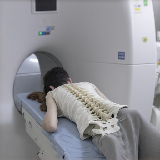
During my appointment, I learned that spinal scans typically include MRI, CT, or X-ray imaging, depending on the suspected issue. My doctor explained that these imaging techniques provide detailed views of the spinal bones, discs, nerves, and surrounding tissues, which are vital for accurate diagnosis. I was surprised by how comfortable the procedure was—no invasive steps involved, just lying still while the machine captured images.
The Role of MRI and CT in Orthopedic Diagnostics
I found it fascinating that MRI scans are excellent for soft tissue evaluation, revealing herniated discs or nerve impingements, while CT scans give a clearer picture of bone structures. The choice of imaging depends on the clinical questions at hand. This tailored approach ensures that the orthopedic specialist can pinpoint the exact cause of spinal pain or dysfunction.
What to Expect During Your Spinal Scan: Practical Tips
If you’re preparing for a spinal scan, I recommend wearing comfortable clothing and informing your technician of any metal implants or devices. The process itself is quick—mine took about 30 minutes—and you might be asked to hold your breath briefly during certain shots. It’s normal to feel a bit uneasy lying still, but overall, it’s a painless and safe procedure.
Why is Accurate Imaging Critical for Effective Treatment?
From my experience, clear and precise images are the foundation of effective orthopedic care. They allow your doctor to develop a targeted treatment plan, whether that involves physical therapy, injections, or surgery. For instance, I discovered a herniated disc that required specific interventions, which I learned are often guided by the detailed insights from these scans. For more insights, I checked out [this authoritative source](https://www.orthoinfo.org) on diagnostic imaging techniques.
If you’re dealing with chronic back pain or suspect a spinal issue, don’t hesitate to ask your orthopedic specialist about the imaging options available. It’s a crucial step toward understanding your condition and exploring your treatment choices.
Feeling inspired to learn more? Share your experiences with spinal imaging in the comments or explore additional resources for spinal health and orthopedic care [here](https://njorthopedicdoctor.xyz/contact-us).
The Critical Role of High-Resolution Imaging in Accurate Spinal Diagnosis
In the realm of orthopedic spinal care, the precision of diagnosis hinges on the quality of imaging technologies. Advanced MRI and CT scans not only reveal the structural intricacies of the spine but also uncover subtle abnormalities that might elude traditional X-rays. As experts, we recognize that detailed imaging is essential for crafting personalized treatment plans, whether for non-invasive therapies or surgical interventions. For example, a nuanced MRI can differentiate between various types of disc herniations, guiding targeted physical therapy or minimally invasive procedures.
How Do Cutting-Edge Imaging Techniques Enhance Treatment Outcomes?
Emerging imaging modalities in 2025 incorporate functional imaging alongside structural assessments, providing insights into nerve activity and blood flow within spinal tissues. This integration allows for a more comprehensive understanding of pain mechanisms and can predict responses to specific treatments. Moreover, innovations like 3D reconstruction and real-time imaging during interventions have revolutionized surgical precision, reducing operative risks and improving recovery times.
Expert Tips for Patients: Preparing for Your Spinal Imaging Appointment
Patients should communicate any metallic implants, pacemakers, or other devices before imaging, as these can interfere with scan quality. Wearing loose, comfortable clothing without metal fasteners can streamline the process. It’s also advisable to remain still during the scan to ensure crisp images—an aspect often overlooked but crucial for accurate diagnosis. Remember, detailed images pave the way for effective, targeted therapies, emphasizing the importance of choosing a facility equipped with the latest imaging technology.
What Are the Limitations of Current Imaging, and How Are Experts Overcoming Them?
While advanced imaging has significantly improved diagnostic accuracy, it is not without limitations. For instance, distinguishing between scar tissue and recurrent disc herniation can still pose challenges. To address this, experts are increasingly combining imaging results with clinical assessments and nerve conduction studies. Additionally, ongoing research into molecular imaging aims to visualize inflammatory processes at the cellular level, promising even greater diagnostic precision in the future. Staying informed about these innovations helps both clinicians and patients make better decisions—visit this trusted source for in-depth insights into diagnostic advancements.
Interested in exploring how imaging choices influence treatment strategies? Share your questions or experiences below, or consider reading more about top spine specialists in 2025 to find the right expert for your needs.
Beyond the Surface: The Nuanced World of Spinal Imaging and Its Impact on Treatment
Reflecting on my journey through advanced spinal imaging, I realize how much these technological marvels have transformed my understanding of spinal health. It’s not just about getting clear pictures; it’s about the stories these images tell—stories that reveal subtle abnormalities, early signs of degeneration, or even tiny nerve impingements that might otherwise go unnoticed. Such detailed insights empower both patients and doctors to craft truly personalized treatment plans, moving beyond one-size-fits-all approaches.
What Are the Hidden Layers in Spinal Imaging That Make a Difference?
In my experience, the real magic lies in the ability of high-resolution MRI and 3D reconstructions to uncover layers of information that traditional X-rays miss. For example, a delicate herniation or minor disc bulge might seem insignificant on a standard scan but could be the key to understanding persistent symptoms. Advanced imaging techniques also allow for the visualization of nerve root inflammation or microvascular changes, which are crucial for diagnosing complex cases. These nuances are what differentiate effective treatment from trial and error, and I’ve seen firsthand how they shape my path to recovery.
How Do Cutting-Edge Techniques Evolve and Personalize Treatment?
Emerging modalities like functional MRI, which assess nerve activity, and real-time intraoperative imaging have added new dimensions to spinal diagnostics. They enable surgeons to perform minimally invasive procedures with pinpoint accuracy, significantly reducing recovery times and improving outcomes. I’ve learned that staying informed about these innovations isn’t just for specialists; it’s vital for patients seeking the best care. For instance, integrating real-time imaging during interventions can help tailor surgical approaches to the patient’s unique anatomy, a concept I find both fascinating and reassuring. For those interested, exploring how these technologies are shaping the future, as discussed in recent studies, can provide valuable perspective.
What Are the Practical Considerations for Patients Preparing for Advanced Spinal Imaging?
From my personal experience, preparation is key. Wearing comfortable clothing, informing your technician about any implants, and remaining still during scans ensure the best quality images. Additionally, understanding the limitations—such as the inability to distinguish scar tissue from recurrent herniation—helps manage expectations. It’s also worth discussing with your doctor whether newer techniques like diffusion tensor imaging or molecular imaging might be suitable for your case. These advancements are pushing the boundaries of what’s possible in diagnostics and treatment planning. For more detailed guidance, visiting trusted sources like this authoritative site can be very helpful.
If you’ve experienced similar journeys or have questions about the latest in spinal imaging, I warmly invite you to share your thoughts in the comments. Connecting with others who are navigating these complex decisions makes the journey more meaningful and less daunting.
Future Horizons: The Promise of Molecular and Functional Imaging
Looking ahead, the integration of molecular imaging and functional assessments promises to revolutionize spinal diagnostics even further. Imagine being able to see inflammatory processes at the cellular level or predict how your nerve tissues will respond to specific therapies—these are no longer distant dreams but emerging realities. Such technologies could drastically reduce misdiagnoses and enable truly personalized medicine, aligning perfectly with my belief that understanding the patient’s unique biological landscape is essential for effective care. To stay updated on these exciting developments, I recommend following recent publications and innovations shared by leading orthopedic research centers.
Unlocking the Next Frontier in Spinal Imaging: Molecular and Functional Insights
As a seasoned orthopedic professional, I am continually fascinated by how cutting-edge imaging technologies redefine our diagnostic capabilities. Traditional MRI and CT scans have long been the cornerstone of spinal assessment, providing detailed structural information that guides effective treatment. However, recent advances in molecular and functional imaging are opening new horizons, enabling us to visualize biological processes at a cellular level and understand nerve activity in real time. This evolution transforms our approach from merely observing anatomical anomalies to comprehending the underlying pathophysiology driving pain and dysfunction.
How Do Molecular Imaging Techniques Enhance Spinal Diagnosis?
Molecular imaging, such as positron emission tomography (PET) combined with MRI, allows us to detect inflammatory markers, metabolic activity, and cellular changes associated with degenerative disc disease, spondylitis, or nerve inflammation. For instance, by visualizing inflammatory cytokines in the spinal tissues, clinicians can identify active disease processes that might not be apparent on conventional scans. This precision helps tailor personalized treatment plans, whether opting for conservative management or surgical intervention. An insightful resource on these innovations can be found at this authoritative source. As practitioners, integrating molecular insights enhances our diagnostic confidence and guides targeted therapies, ultimately improving patient outcomes.

Furthermore, advancements in molecular imaging contribute to early detection, allowing interventions before significant structural damage occurs, which is crucial for conditions like disc degeneration or facet joint inflammation. This proactive approach aligns with the growing emphasis on preventive orthopedics and personalized medicine.
What Role Does Functional MRI Play in Understanding Nerve Activity?
Functional MRI (fMRI) measures changes in blood flow related to nerve activity, providing dynamic insights into how pain signals originate and propagate within the spinal cord and nerve roots. This technology is invaluable in complex cases where structural imaging alone does not explain persistent symptoms. For example, fMRI can reveal hyperactive nerve pathways in patients with radiculopathy or neuropathic pain, informing more precise interventions such as nerve blocks or minimally invasive surgeries.
Incorporating functional assessments into our diagnostic repertoire is revolutionary, enabling us to move beyond static images and into a realm where we understand the biological and neurological underpinnings of spinal pain. This comprehensive perspective fosters a more holistic treatment approach, combining structural correction with neuromodulation strategies. To stay at the forefront, I recommend following recent publications from leading research centers specializing in orthopedic imaging advancements.
How Will These Innovations Shape Future Treatment Paradigms?
The integration of molecular and functional imaging will undoubtedly influence the development of targeted therapies, including biologic injections, regenerative medicine, and neuromodulation techniques. For example, identifying active inflammation at the cellular level can guide the use of biologics, while understanding nerve hyperactivity can optimize nerve ablation procedures. These personalized strategies promise to reduce unnecessary interventions and enhance recovery trajectories.
Moreover, real-time intraoperative imaging that combines structural and functional data will elevate surgical precision, minimizing tissue damage and improving long-term stability. As these technologies evolve, collaboration between radiologists, orthopedic surgeons, and pain specialists will become increasingly vital to harness their full potential.
Are You Ready to Explore These Future-Forward Diagnostics?
If you’re committed to advancing your understanding of spinal health and optimizing patient care, I invite you to delve deeper into these emerging imaging modalities. Embracing molecular and functional insights not only enhances diagnostic accuracy but also aligns with the broader goals of personalized, minimally invasive treatment. For comprehensive guidance tailored to your clinical practice, consider exploring resources at this trusted site. Sharing your experiences or questions in the comments can foster valuable exchanges as we collectively navigate the future of orthopedic diagnostics and patient care.
Things I Wish I Knew Earlier (or You Might Find Surprising)
Hidden Layers of Detail
One thing I discovered is that advanced imaging techniques like MRI and CT scans reveal subtle abnormalities that traditional X-rays often miss, which can be a game-changer in diagnosing complex spinal issues.
The Power of Personal Experience
My own journey with spinal imaging showed me how understanding these images empowers patients to participate actively in their treatment decisions, making the process less intimidating.
Technological Evolution
I was amazed to learn how emerging technologies such as functional MRI and molecular imaging are pushing the boundaries of diagnostics, offering insights into nerve activity and inflammation at a cellular level.
Preparation Matters
Practical tips like wearing loose clothing and informing your technician about implants can significantly improve scan quality and comfort.
The Limitations and Future of Imaging
While current imaging is incredibly detailed, it still struggles with distinguishing scar tissue from recurrent herniation. Thankfully, ongoing research promises even more precise tools on the horizon.
Resources I’ve Come to Trust Over Time
- OrthoInfo from the AAOS: This site offers comprehensive, peer-reviewed articles that deepen my understanding of diagnostic imaging and its role in orthopedic care.
- National Institutes of Health (NIH): Their research publications provide cutting-edge insights into the latest advances in imaging technology.
- RadiologyInfo.org: A user-friendly resource that explains imaging procedures in accessible language, which I found helpful when preparing for scans.
Parting Thoughts from My Perspective
Reflecting on my experiences, I realize that detailed spinal imaging is not just about getting pictures—it’s about uncovering the stories these images tell, stories that can lead to truly personalized and effective treatment plans. If this resonates with you, I’d love to hear your thoughts. Feel free to share your own experiences or questions in the comments, and remember, staying informed is the first step towards better spinal health and recovery.