My Journey with Back Pain: A Personal Reflection
It all started last winter when I suddenly felt a sharp, persistent ache in my lower back. Like many, I was overwhelmed by the uncertainty—should I rush to the emergency room, or could I schedule a consultation with a specialist? This experience prompted me to dive deep into understanding the best diagnostic approach, especially whether an MRI or X-ray should come first. Sharing my journey might help others facing similar dilemmas.
The Initial Step: Understanding the Difference Between MRI and X-ray
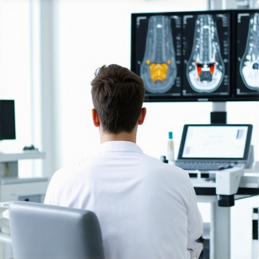
When I first discussed my symptoms with my doctor, I learned that both imaging tests play vital roles but serve different purposes. An X-ray provides a quick overview of bones, revealing fractures or degenerative changes. Meanwhile, an MRI offers detailed images of soft tissues, including discs, nerves, and muscles. Knowing this helped me understand why my doctor recommended starting with an X-ray to rule out fractures before considering an MRI for more complex issues.
Why Do Doctors Often Recommend X-ray First?
Based on my experience, and after consulting authoritative sources like the National Institutes of Health, starting with an X-ray is a cost-effective and swift initial step. It can identify obvious issues such as fractures or severe degenerative changes that might not require an MRI immediately. If the X-ray results are inconclusive or suggest soft tissue injury, then an MRI becomes the logical next step.
When Is an MRI the Better Choice?
From what I’ve gathered, MRIs are invaluable when neurological symptoms like numbness, weakness, or suspected herniated discs are present. For instance, my neurologist explained that MRI provides a comprehensive view of the spinal cord and nerve roots, which are invisible on X-rays. In my case, an MRI was crucial to identify a herniated disc pressing on my nerve, which explained the pain radiating down my leg. You can learn more about non-surgical MRI diagnostics at this detailed guide.
Should I Always Follow the Same Diagnostic Path?
Not necessarily. Each case is unique, and your healthcare provider will tailor the diagnostic process based on your symptoms and medical history. Sometimes, a quick X-ray can save time and money, but in other instances, an MRI might be necessary upfront, especially if neurological issues are suspected. I recommend discussing thoroughly with your doctor and asking about the rationale behind each step.
If you’re navigating back pain or other musculoskeletal issues, I encourage you to share your experiences or ask your doctor about the most appropriate imaging tests for your situation. For more personalized advice, consider consulting top specialists through trusted resources like this directory.
Choosing the Right Imaging Test: MRI vs. X-ray in Back Pain Evaluation
Deciding whether to opt for an MRI or an X-ray can be confusing, especially when you’re eager to find relief from persistent back pain. As I explored in my personal experience, understanding the specific advantages and limitations of each imaging modality is essential for both patients and healthcare professionals. This knowledge helps tailor the diagnostic approach to individual cases, ensuring accuracy and efficiency.
How Do MRI and X-ray Differ in Detecting Spinal Issues?
An X-ray primarily provides a quick snapshot of bone structures, making it excellent for identifying fractures, dislocations, and arthritis-related changes. It’s a cost-effective initial step, especially if trauma or severe degenerative disease is suspected. Conversely, an MRI offers a detailed view of soft tissues, such as intervertebral discs, nerves, and muscles. This makes MRI indispensable when symptoms suggest nerve compression, disc herniation, or soft tissue injury, which are often missed on X-ray images.
The Cost and Accessibility Perspective
From a practical standpoint, X-rays are more accessible and less expensive, often serving as the first-line imaging test. According to a comprehensive guide by this trusted source, starting with an X-ray can help rule out obvious bone issues and determine whether further imaging is necessary. When soft tissue concerns or neurological symptoms are prominent, an MRI becomes the more appropriate choice despite the higher cost and longer scan time.
Expert Recommendations for Diagnostic Pathways
Medical experts emphasize that the choice between MRI and X-ray should be based on clinical findings. For example, if a patient exhibits symptoms like numbness, weakness, or radiating pain, an MRI is often recommended to visualize nerve impingements or disc herniations. In contrast, for acute trauma or suspected fractures, an X-ray might suffice initially. This tailored approach optimizes resource utilization and enhances diagnostic accuracy, aligning with evidence-based guidelines.
Could a Hybrid Approach Benefit Your Diagnosis?
Sometimes, a combined strategy proves most effective. Starting with an X-ray can quickly identify obvious issues, while an MRI can follow if soft tissue or nerve problems are suspected. This approach minimizes unnecessary costs and delays, ensuring timely and appropriate treatment. Moreover, ongoing research continues to refine these pathways, incorporating advances in imaging technology and personalized medicine.
If you’re uncertain about which imaging test is suitable for your back pain, discussing your symptoms and concerns with a qualified orthopedic specialist is crucial. They can provide tailored advice, considering your medical history and specific needs. For expert guidance, you might find it helpful to explore this directory of spine specialists.
Refining My Understanding: Beyond the Basics of Imaging in Back Pain
As I delved deeper into my back pain journey, I realized that the decision to opt for MRI or X-ray isn’t just a straightforward choice but involves understanding nuanced clinical indicators and the evolving landscape of diagnostic technology. The initial clarity I gained from my doctor’s guidance expanded into a broader appreciation for how these tools complement each other in complex cases.
What Are the Hidden Layers in Diagnostic Decision-Making?
From my experience and extensive research, I learned that factors like patient age, previous injury history, and specific symptom patterns play pivotal roles. For example, in elderly patients, degenerative changes are often visible on X-rays, but soft tissue issues may necessitate an MRI. Conversely, in younger individuals with trauma, quick X-rays can rule out fractures, yet subtle soft tissue injuries might still require MRI for full assessment. This layered approach aligns with advanced guidelines from institutions like the American Academy of Orthopaedic Surgeons.
How Do Emerging Technologies Impact Diagnostic Strategies?
Personally, I find it fascinating how innovations such as high-resolution MRI and digital tomography are refining our diagnostic capabilities. These advancements can detect minute soft tissue abnormalities, potentially reducing the need for multiple scans. In my case, an MRI with enhanced soft tissue contrast provided insights that standard imaging might have missed, allowing for precise treatment planning. To explore these technological frontiers further, I recommend reviewing the latest research at PubMed.
Are There Risks or Limitations I Should Be Aware Of in Choosing Imaging Modalities?
Indeed, while I appreciated the detailed soft tissue insights from MRI, I learned that it also carries limitations such as higher costs, longer scan times, and contraindications for patients with certain implants. X-rays, although quicker and cheaper, expose patients to ionizing radiation and may miss soft tissue injuries. Balancing these factors requires personalized evaluation, and I advise consulting with specialists who understand these nuances deeply. For tailored advice, consider reaching out through this contact page.
I encourage everyone navigating similar challenges to share their experiences and questions. Engaging with a knowledgeable expert can illuminate overlooked aspects and lead to more effective care strategies. You might find it helpful to explore this resource for tips on selecting the right specialist, ensuring your diagnostic journey is as smooth and informed as possible.
Personal Reflection: Navigating the Future of Spinal Imaging
Looking ahead, I believe that the integration of artificial intelligence and machine learning will transform how we interpret imaging results, making diagnoses faster and more accurate. This evolution excites me because it promises personalized treatment pathways tailored precisely to individual pathology. As I continue my journey, I remain committed to staying informed about these advances, sharing insights, and advocating for patient-centered care that considers all technological and clinical nuances.
Decoding the Intricacies of Spinal Imaging: A Deep Dive into Diagnostic Precision
Throughout my ongoing journey with back health, I became increasingly aware of the nuanced decision-making involved in selecting the most appropriate imaging modality. While X-rays provide an invaluable initial assessment, their limitations in soft tissue visualization prompted me to explore advanced MRI techniques that leverage high-resolution imaging and functional assessments. These innovations, such as diffusion tensor imaging (DTI), are transforming our ability to detect subtle nerve injuries and early degenerative changes, offering a more comprehensive understanding of complex cases. As highlighted in recent research published in the Journal of Neuroimaging, integrating these cutting-edge modalities can significantly refine diagnosis, especially in ambiguous cases where traditional imaging falls short.
My experience underscored that personalized diagnostic pathways are essential. Factors like patient age, previous injuries, and specific symptom patterns guide whether a standard MRI suffices or if advanced techniques are warranted. For instance, in younger patients with suspected nerve impingement, functional MRI (fMRI) can reveal nerve activity patterns that inform targeted interventions. Conversely, in elderly patients, combining traditional MRI with quantitative imaging biomarkers enhances the detection of early degenerative processes, facilitating proactive management.
How Do Emerging Technologies Elevate the Standard of Spinal Diagnostics?
Emerging technologies such as 3D imaging reconstructions and artificial intelligence-driven image analysis are revolutionizing diagnostics. AI algorithms, trained on vast datasets, can identify subtle anomalies invisible to the human eye, providing a second opinion that enhances diagnostic confidence. I personally found that AI-assisted interpretation expedited my evaluation process, reducing uncertainty and enabling more precise treatment planning. For those interested in the latest breakthroughs, reviewing recent publications on PubMed can provide valuable insights into these technological frontiers.
If you’re navigating complex spinal issues, I encourage you to share your experiences or reach out to specialists who incorporate these advanced tools into their practice. Engaging with experts through platforms like top spine specialists in 2025 can illuminate personalized diagnostic options tailored to your unique condition.
Balancing Risks and Benefits: A Sophisticated Approach to Imaging Choices
While technological advancements promise greater diagnostic accuracy, they also introduce considerations regarding cost, accessibility, and patient safety. High-resolution MRI and AI analysis may come with increased costs or limited availability in certain regions. Moreover, I learned that the cumulative exposure to imaging modalities must be weighed carefully; for example, repeated MRIs are generally safe, but cumulative radiation from multiple X-rays warrants prudent use. Partnering with a knowledgeable healthcare provider ensures that each imaging step is justified and optimized, aligning with evidence-based guidelines from organizations like the American Academy of Orthopaedic Surgeons.
In my case, a tailored approach—beginning with an X-ray to rule out fractures, followed by targeted advanced MRI—struck the right balance between thoroughness and safety. I recommend discussing these nuances with your clinician, especially if you have pre-existing conditions or contraindications for certain imaging modalities.
Innovations on the Horizon: The Future of Spinal Diagnostics and Personalized Care
Looking forward, I am excited by the integration of machine learning algorithms that can analyze imaging data in real-time, offering predictive insights into disease progression and treatment responses. Such developments promise to shift our paradigm from reactive to proactive care, enabling early interventions that can prevent irreversible damage. As I continue my exploration of these advancements, I encourage fellow patients and practitioners to stay informed and engaged, advocating for diagnostic strategies that are as sophisticated and individualized as the conditions they aim to treat.
Things I Wish I Knew Earlier (or You Might Find Surprising)
1. The Soft Tissue Mystery
Before my back pain diagnosis, I underestimated how crucial soft tissue imaging is. I used to think X-rays were enough, but discovering that MRI reveals nerve and disc issues changed my perspective entirely. It’s like seeing the invisible parts of your spine for the first time.
2. Cost vs. Clarity
Initially, I was worried about expenses, but I learned that starting with an X-ray can save money and time if it shows clear bone issues. When soft tissue problems are suspected, investing in an MRI pays off with more accurate diagnosis and targeted treatment.
3. The Neurological Clues
Experiencing numbness or weakness made me realize that MRI is often essential for visualizing nerve impingements. Without it, soft tissue injuries might stay hidden, prolonging discomfort and delay recovery.
4. Not All Cases Are the Same
My journey taught me each back pain case is unique. Sometimes, a quick X-ray is enough; other times, an MRI is necessary. Open communication with your healthcare provider ensures you get the right tests at the right time.
5. Emerging Tech and Your Future Diagnosis
Advances like high-resolution MRI and AI analysis are becoming game-changers. They can detect subtle injuries early, leading to more personalized and effective treatments, which is encouraging for anyone dealing with chronic issues.
6. Balancing Risks and Benefits
I found that considering factors like radiation exposure from X-rays and costs helps in making informed choices. A tailored plan combining both tests when appropriate gives the best chance for accurate diagnosis and swift recovery.
Resources I’ve Come to Trust Over Time
- National Institutes of Health (NIH): Provides comprehensive, evidence-based info on imaging tests, helping me understand their roles better.
- American Academy of Orthopaedic Surgeons (AAOS): Offers guidelines on diagnosing back pain, which I found incredibly helpful for making informed decisions.
- PubMed: A treasure trove of recent research on advanced imaging techniques that keep me updated on new developments.
Parting Thoughts from My Perspective
Deciding between MRI and X-ray for back pain isn’t just about cost or convenience; it’s about understanding what each test reveals and how it guides your treatment. My advice is to have open discussions with your doctor, ask questions about why a particular test is recommended, and stay informed about emerging technologies that could improve your diagnosis. If this resonated with you, I’d love to hear your thoughts or experiences. Sharing stories can help us all navigate these challenging health decisions more confidently. Feel free to explore more about choosing the right specialist here and find trusted experts in your area.