Why Precision Matters: The Backbone of Orthopedic Diagnostic Imaging in NJ
Orthopedic diagnostic imaging is a pivotal step in diagnosing and managing musculoskeletal disorders, especially in a state like New Jersey where active lifestyles and aging populations converge. These imaging services provide clinicians with a window into the complexities of bones, joints, muscles, and connective tissues, enabling tailored treatment plans that optimize recovery and mobility.
From subtle fractures to chronic degenerative diseases, high-quality imaging ensures no detail is overlooked. This precision is not just about technology but about enhancing patient outcomes through early detection and accurate assessment.
Revealing the Spectrum: Key Imaging Modalities Transforming Orthopedic Care
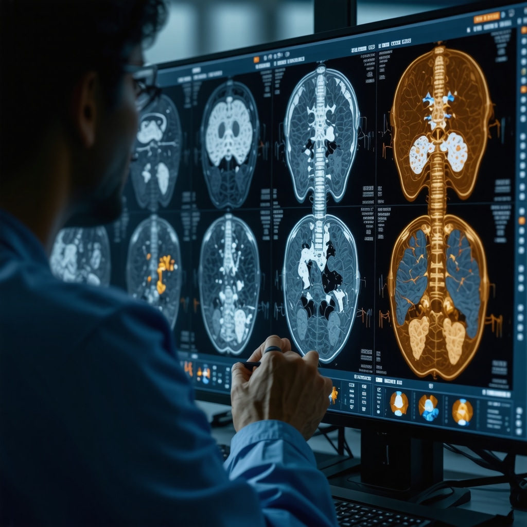
In NJ, orthopedic specialists rely on a suite of advanced imaging techniques including X-rays, MRI (Magnetic Resonance Imaging), CT (Computed Tomography), and ultrasound. Each modality serves unique diagnostic purposes. For example, X-rays remain the gold standard for detecting bone fractures and alignment issues, while MRI excels in visualizing soft tissue injuries such as ligament tears or cartilage damage without radiation exposure.
CT scans provide detailed cross-sectional images, crucial for complex fractures or preoperative planning. Ultrasound offers a real-time, dynamic view of tendons and joints and is often used for guided injections. This diverse arsenal ensures comprehensive evaluation tailored to each patient’s clinical presentation.
How Do Orthopedic Imaging Services Enhance Treatment Accuracy and Speed?
Orthopedic imaging services accelerate diagnosis by revealing the underlying pathology that might be missed in physical examinations alone. For instance, an MRI can detect early cartilage degradation before symptoms worsen, enabling interventions that may delay or prevent invasive surgery. These imaging insights allow orthopedic surgeons and clinicians in NJ to formulate precise treatment strategies, whether surgical or conservative, improving recovery timelines and reducing complications.
Moreover, the integration of imaging with electronic health records facilitates multidisciplinary collaboration, a critical factor in managing complex orthopedic cases.
Experience Meets Innovation: Navigating NJ’s Orthopedic Imaging Landscape
New Jersey’s orthopedic centers combine cutting-edge imaging technologies with seasoned radiologists and orthopedic experts who interpret results within the broader clinical context. This synergy ensures that diagnostic imaging is not merely a technical procedure but an integral part of personalized patient care.
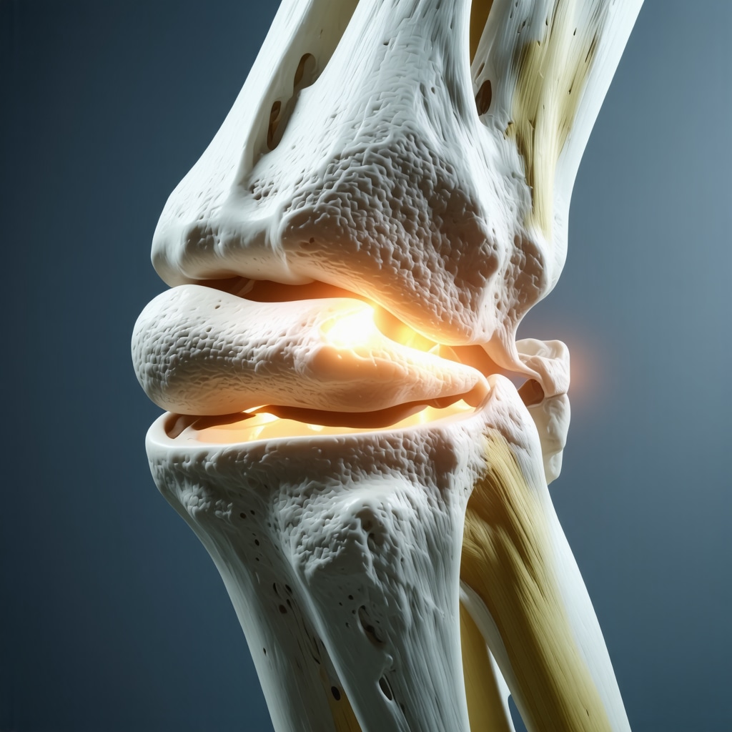
Consider a middle-aged athlete presenting with persistent knee pain. Advanced MRI imaging could reveal a meniscus tear that standard X-rays would miss, guiding timely arthroscopic repair. Such practical case scenarios underline the indispensable role of comprehensive imaging services in NJ’s orthopedic care continuum.
Empowering Patients: What to Expect During Your Orthopedic Imaging Appointment in NJ
Understanding the process can alleviate patient anxiety. Appointments typically begin with a clinical evaluation followed by the imaging procedure that may vary in duration and preparation. For example, MRI requires patients to remain still inside a scanner for 30-60 minutes, while X-rays are quicker but sometimes require multiple views.
Technologists in NJ orthopedic imaging centers prioritize patient comfort and safety, adhering to strict protocols to minimize radiation exposure and optimize image quality. Clear communication about the procedure and follow-up steps fosters transparency and trust.
For those interested in detailed insights on orthopedic appointments, exploring what to expect during your visit can be very helpful.
Elevate Your Orthopedic Care Journey: Engage with NJ’s Imaging Experts
If you or a loved one are navigating orthopedic challenges, tapping into comprehensive diagnostic imaging services in NJ can be a game-changer. These services not only clarify diagnosis but also empower your healthcare team to design effective, individualized treatments.
Curious about how specialized imaging can improve your orthopedic outcomes? Feel free to contact NJ’s orthopedic imaging specialists to discuss your needs and explore the best diagnostic options available.
Explore more on orthopedic diagnostic innovations and stay informed about treatment options by visiting trusted sources like the American Academy of Orthopaedic Surgeons, a leading authority in musculoskeletal health.
Personal Reflections on Choosing the Right Imaging Modality
When I first faced persistent back pain, the sheer variety of imaging options felt overwhelming. I vividly remember my initial appointment with an orthopedic specialist in NJ, where the discussion quickly turned to whether an MRI, CT scan, or X-ray would best reveal the cause. The doctor patiently explained that while X-rays are excellent for bone fractures, MRI is superior for soft tissue issues like herniated discs or ligament tears. This clarity helped me appreciate how tailored imaging leads to better outcomes.
For those curious about selecting the right imaging, checking out how to choose the right orthopedic surgeon for your spine can offer additional guidance on the interplay between diagnosis and treatment.
Integrating Imaging with Rehabilitation: A Personal Journey
After my diagnosis, imaging didn’t just stop at identifying the problem—it continued to guide my recovery. The detailed MRI results allowed my orthopedic team to design a rehabilitation program that focused on strengthening the affected area while avoiding further injury. I found myself frequently consulting the images alongside my physical therapist, which made the recovery process more transparent and motivating.
If you or someone you know is recovering from surgery, exploring orthopedic rehab tips after lumbar fusion surgery can provide practical strategies that complement imaging insights.
How Can Patients Be More Proactive in Understanding Their Imaging Results?
One question I often ponder is how patients can demystify their imaging reports to actively participate in treatment decisions. From my experience and conversations with NJ orthopedic experts, asking your doctor to walk you through the images in simple terms can make a huge difference. Sometimes, even bringing a trusted family member or friend to appointments helps in retaining details. Digital platforms now often allow access to your imaging results, so reviewing them at home with additional resources can deepen your understanding.
Trusting the Experts and Staying Informed
In NJ, the combination of experienced radiologists and orthopedic surgeons ensures that imaging interpretations are accurate and clinically relevant. According to the American Academy of Orthopaedic Surgeons, having access to high-quality imaging services is essential for timely interventions and effective treatment plans (AAOS).
Personally, knowing that such expertise is available locally gave me peace of mind during my treatment journey. If you’re seeking specialized care, resources like top orthopedic spine specialists to trust in 2025 can be a great starting point.
Have you had experiences with orthopedic imaging that changed how you viewed your treatment? Share your story in the comments below or explore more about effective non-surgical care for herniated discs to learn how imaging supports various treatment pathways.
Harnessing 3D Imaging and AI: The Next Frontier in Orthopedic Diagnostics
New Jersey’s orthopedic imaging landscape is rapidly evolving with the integration of three-dimensional (3D) imaging technologies and artificial intelligence (AI) algorithms. These advancements are reshaping how clinicians visualize and interpret complex musculoskeletal conditions, allowing for unprecedented precision in diagnosis and treatment planning.
3D imaging, particularly through techniques like cone-beam computed tomography (CBCT) and advanced CT reconstructions, provides volumetric views of bone and joint structures that traditional two-dimensional X-rays cannot offer. This dimensional depth facilitates better assessment of fracture morphology, subtle deformities, and post-surgical outcomes.
Simultaneously, AI-driven image analysis tools are enhancing radiologist efficiency and diagnostic accuracy by automatically detecting anomalies, quantifying tissue degeneration, and even predicting disease progression trajectories. For example, AI algorithms trained on large datasets can identify early osteoarthritis changes on MRI scans before they become clinically apparent, enabling proactive interventions.
What Are the Clinical Benefits and Limitations of AI-Powered Orthopedic Imaging?
While AI integration holds immense promise, understanding its clinical impact requires balancing benefits with current limitations. On the plus side, AI can reduce inter-observer variability by standardizing image interpretation and flagging subtle pathologies that might be overlooked in busy clinical settings. This ensures that NJ patients receive earlier and more accurate diagnoses, which is critical for conditions like stress fractures or early cartilage wear.
However, AI systems depend heavily on the quality and diversity of training data. Biases or gaps in datasets may lead to false positives or missed findings, underscoring the need for expert radiologist oversight. Moreover, the integration of AI tools into existing clinical workflows requires careful validation and regulatory approvals, which can delay widespread adoption.
Despite these challenges, the American Journal of Roentgenology highlights that AI-assisted imaging is already transforming musculoskeletal radiology by complementing human expertise rather than replacing it (American Journal of Roentgenology, 2020).
Precision-Guided Interventions: Leveraging Imaging for Therapeutic Excellence
Beyond diagnosis, advanced orthopedic imaging in NJ facilitates precision-guided interventions, such as ultrasound-guided joint injections and CT-navigated minimally invasive surgeries. These techniques improve targeting accuracy, reduce procedural complications, and enhance patient comfort.
For instance, using dynamic ultrasound to guide corticosteroid injections in the shoulder or knee ensures medication delivery directly into the inflamed synovial space, maximizing efficacy. Similarly, intraoperative CT scans allow orthopedic surgeons to verify implant positioning in real-time during complex spinal surgeries, reducing the need for revision procedures.
Such integration of imaging and intervention epitomizes personalized medicine, where diagnostic clarity seamlessly transitions into therapeutic action.

Empowering Patients Through Imaging Literacy: Strategies for Enhanced Engagement
Patients often face challenges interpreting complex imaging reports and understanding their implications. Empowering patients with imaging literacy is crucial for shared decision-making and adherence to treatment plans.
Clinicians in NJ are adopting strategies such as visual aids, annotated imaging reviews during consultations, and digital portals that provide accessible explanations alongside radiology reports. Encouraging patients to ask targeted questions about their images fosters a collaborative environment and demystifies the diagnostic process.
Moreover, educational resources tailored to orthopedic imaging can help patients grasp the significance of findings like ligament tears, cartilage thinning, or bone edema, enabling them to actively participate in their recovery journey.
Interested in deepening your understanding of orthopedic imaging? Connect with NJ’s expert radiologists and orthopedic specialists who can guide you through your diagnostic journey with clarity and expertise.
Harnessing Cutting-Edge Orthopedic Imaging Technologies in New Jersey
Building on New Jersey’s robust orthopedic imaging infrastructure, the advent of 3D imaging and artificial intelligence (AI) is revolutionizing musculoskeletal diagnostics. These innovations are not merely incremental upgrades but transformative tools that deepen clinical insights and personalize patient care pathways.
3D imaging modalities, including cone-beam computed tomography (CBCT) and volumetric CT reconstructions, offer unprecedented spatial visualization of complex bone structures, essential for nuanced fracture assessment and surgical planning. By transcending traditional two-dimensional limitations, clinicians gain a holistic understanding of anatomical relationships, enhancing diagnostic precision.
Simultaneously, AI algorithms trained on extensive clinical datasets augment radiologist expertise by automating anomaly detection, quantifying degenerative changes, and prognosticating disease trajectories. Such capabilities enable earlier intervention strategies, particularly in degenerative joint diseases and subtle stress fractures that challenge conventional imaging interpretation.
What Are the Clinical Benefits and Limitations of AI-Powered Orthopedic Imaging?
While AI integration promises enhanced diagnostic accuracy and workflow efficiency, it necessitates careful scrutiny of its limitations. Notably, AI’s performance is contingent upon the quality and representativeness of its training data; biases can precipitate false positives or negatives, underscoring the indispensability of expert radiologist oversight to contextualize findings. Furthermore, regulatory hurdles and the need for seamless clinical workflow integration remain challenges to its widespread adoption.
According to a 2020 study published in the American Journal of Roentgenology, AI serves as a complementary adjunct to human interpretation rather than a replacement, amplifying diagnostic confidence and consistency in musculoskeletal radiology.
Precision-Guided Therapeutic Interventions: The Symbiosis of Imaging and Treatment
Orthopedic imaging’s utility extends beyond diagnostics into the realm of precision-guided therapeutics. In New Jersey, ultrasound-guided injections and CT-navigated minimally invasive surgeries exemplify how imaging facilitates targeted interventions that optimize efficacy and minimize iatrogenic risks.
Dynamic ultrasound enables real-time visualization during corticosteroid or platelet-rich plasma (PRP) injections, ensuring medication delivery directly into pathological tissues such as inflamed synovium or degenerated tendons. Similarly, intraoperative CT imaging provides immediate feedback on implant positioning during spinal instrumentation, mitigating revision rates and enhancing surgical outcomes.
This integrated approach epitomizes personalized medicine, where diagnostic acuity seamlessly informs and improves therapeutic precision.

Empowering Patients Through Enhanced Imaging Literacy: Strategies for Shared Decision-Making
Despite the sophistication of imaging modalities, patients frequently encounter difficulties interpreting complex radiological reports, which may hinder informed participation in their care. Recognizing this, New Jersey orthopedic centers are pioneering patient-centered educational initiatives to elevate imaging literacy.
These include the use of annotated imaging during consultations, accessible digital portals offering simplified explanations, and encouraging proactive patient inquiries. Such strategies demystify diagnostic findings—be it ligamentous tears, cartilage attrition, or bone marrow edema—thereby fostering collaborative decision-making and adherence to therapeutic regimens.
Engaging with expert radiologists and orthopedic specialists in NJ can transform the imaging experience from one of uncertainty to empowerment, ultimately enhancing clinical outcomes and patient satisfaction.
Ready to advance your orthopedic care with the latest imaging innovations and expert guidance? Connect with New Jersey’s top imaging specialists today and take an active role in your musculoskeletal health journey.
Frequently Asked Questions (FAQ)
What types of orthopedic imaging are most commonly used in New Jersey?
The primary imaging modalities used include X-rays for bone fractures and alignment, MRI for soft tissue evaluation like ligaments and cartilage, CT scans for detailed cross-sectional bone imaging, and ultrasound for dynamic assessment and guided interventions. Each modality is selected based on the clinical question to provide optimal diagnostic clarity.
How does 3D imaging improve orthopedic diagnosis compared to traditional methods?
3D imaging, such as cone-beam CT and volumetric CT reconstructions, provides volumetric views allowing clinicians to assess complex fractures, deformities, and post-surgical anatomy with greater spatial accuracy. This depth facilitates more precise surgical planning and better understanding of structural relationships than conventional two-dimensional X-rays.
What role does artificial intelligence (AI) play in orthopedic imaging?
AI assists by automating anomaly detection, quantifying degenerative changes, and predicting disease progression. It enhances diagnostic accuracy and efficiency by complementing radiologist expertise, reducing variability, and flagging subtle pathologies that might otherwise be missed, although expert oversight remains essential.
Are there any risks associated with orthopedic imaging procedures?
Most imaging modalities are safe; however, X-rays and CT scans involve exposure to ionizing radiation, which is minimized through strict protocols. MRI and ultrasound do not use radiation but may have contraindications such as implanted medical devices for MRI. Patient safety and comfort are prioritized at New Jersey imaging centers.
How can patients better understand their imaging results?
Patients are encouraged to ask their healthcare providers for simplified explanations and annotated image reviews during consultations. Utilizing accessible digital portals and educational materials can also improve comprehension, empowering patients to actively participate in treatment decisions.
Can imaging guide treatment beyond diagnosis?
Absolutely. Advanced imaging supports precision-guided interventions like ultrasound-guided injections and intraoperative CT for surgical navigation, enhancing treatment accuracy and outcomes by ensuring targeted therapy delivery and implant positioning.
How do orthopedic imaging services integrate with rehabilitation?
Imaging not only diagnoses injuries but also informs rehabilitation by monitoring healing progress and guiding therapy adjustments. Clinicians and therapists can tailor rehabilitation plans based on imaging insights to optimize recovery and prevent re-injury.
What should patients expect during an orthopedic imaging appointment?
Appointments typically start with a clinical evaluation followed by the imaging procedure, which varies in duration and preparation. Technologists focus on patient comfort and safety, providing clear instructions and minimizing radiation exposure when applicable.
How is imaging literacy being improved among patients in New Jersey?
Centers are adopting patient-centered educational initiatives including annotated image reviews, simplified radiology reports, digital portals, and encouraging questions. These approaches help patients understand findings and engage in shared decision-making.
Is AI likely to replace radiologists in orthopedic imaging?
No. AI serves as a powerful adjunct to human expertise by enhancing accuracy and efficiency. Radiologists remain essential for interpreting complex clinical contexts, validating AI findings, and ensuring comprehensive patient care.
Trusted External Sources
- American Academy of Orthopaedic Surgeons (AAOS) – A leading authority providing evidence-based guidelines, educational resources, and updates on orthopedic imaging and treatment advancements.
- American Journal of Roentgenology (AJR) – Offers peer-reviewed research articles on cutting-edge imaging technologies and AI applications in musculoskeletal radiology, underpinning clinical best practices.
- Radiological Society of North America (RSNA) – A premier organization delivering comprehensive resources on radiologic techniques, innovations, and standards relevant to orthopedic imaging.
- National Institute of Arthritis and Musculoskeletal and Skin Diseases (NIAMS) – Provides patient-centered information on musculoskeletal conditions and the role of diagnostic imaging in disease management.
- New Jersey Orthopedic Centers and Academic Hospitals – Institutions such as Rutgers Robert Wood Johnson Medical School contribute to regional expertise, research, and clinical excellence in orthopedic imaging.
Conclusion
Orthopedic diagnostic imaging in New Jersey stands at the confluence of advanced technology, expert interpretation, and patient-centered care. From traditional X-rays to transformative 3D imaging and AI-enhanced analyses, these tools enable precise visualization of musculoskeletal conditions, facilitating tailored treatment and improved outcomes. The integration of imaging into therapeutic interventions and rehabilitation further exemplifies its critical role in comprehensive orthopedic management.
Empowering patients through imaging literacy fosters shared decision-making and enhances treatment adherence, while ongoing innovations promise to refine diagnostic capabilities further. Engaging with New Jersey’s specialized imaging experts offers invaluable support in navigating orthopedic challenges with confidence and clarity.
Take the next step in your orthopedic health journey—explore expert imaging services, ask your healthcare providers insightful questions, and share your experiences to help others benefit from advanced musculoskeletal care.

Reading about the precision required in orthopedic diagnostic imaging in NJ really highlights how essential it is to get the right imaging modality based on the patient’s specific symptoms. I’ve had a family member who struggled with knee pain for months, and initial X-rays didn’t show the full extent of the damage. It was only after an MRI that a meniscus tear was identified, much like the example mentioned in the post. This early detection made a huge difference in avoiding further injury and customizing physical therapy.
What stood out to me is how integrating imaging with electronic health records facilitates better collaboration between specialists, which is so important in complex cases. In my experience, having all diagnostic information readily accessible to both surgeons and therapists led to a more coordinated and effective recovery plan.
I’m curious, for those who have gone through orthopedic imaging in NJ, how did your care team involve you in understanding your imaging results? Did they use annotated images or digital portals to explain the findings? From what I’ve seen, improving imaging literacy can really empower patients to take charge of their recovery. Would love to hear your strategies or suggestions on making this process less intimidating and more interactive for patients.
Building on Jessica Marlowe’s thoughtful observation about the role of imaging integration and patient involvement, I wanted to share my own experience with orthopedic imaging in NJ, which might offer additional perspective on empowering patients. In my case, after a sports injury, my orthopedic care team made a point to review my MRI scans with me in detail, using annotated images displayed on a screen during the consultation. This visual aid made it much easier to understand the nature and extent of my ligament damage, beyond the usual medical jargon.
Moreover, they encouraged me to access my imaging results through a secure digital portal, which included layman-friendly explanations and links to trusted educational resources. This approach gave me a sense of control and reduced uncertainty, allowing me to ask more informed questions throughout my treatment and rehab.
I think one challenge I’ve noticed, however, is that not all clinics implement these strategies consistently, which can leave some patients feeling overwhelmed by complex reports. It makes me wonder: how can NJ orthopedic centers standardize imaging literacy tools to ensure equitable patient engagement? Perhaps peer support groups or virtual workshops could complement digital initiatives, providing collective learning opportunities.
Has anyone else encountered innovative ways their care teams helped demystify imaging results? Sharing these could help foster best practices in patient-centered orthopedic care.
Responding to the insightful points raised by Jessica and Liam about imaging literacy in NJ orthopedic care, I’d like to share how my experience highlighted the critical role of communication throughout the imaging process. During my evaluation for chronic shoulder pain, the radiologist not only reviewed the MRI images with me during the consultation but also provided annotated printouts that I could take home. This really helped me visualize the problem areas, especially the partial rotator cuff tear that was initially hard to grasp.
Additionally, my care team used a patient portal where I could securely review my imaging reports and supplementary educational videos tailored to my condition. I found this combination of verbal explanation, visual aids, and digital resources invaluable in transforming what could be an overwhelming, technical experience into an empowering one.
Given that not all centers adopt these practices uniformly, I agree with Liam’s suggestion about peer support groups and virtual workshops. They could foster a community where patients share insights on navigating orthopedic imaging effectively. From your experience, have you noticed whether these patient-centered educational tools correlate with better adherence to treatment or rehabilitation programs? It would be great to hear if others think making imaging literacy a standardized part of orthopedic care could help improve long-term patient outcomes in NJ.
This post does a fantastic job highlighting how diverse imaging modalities play a crucial role in personalized orthopedic care. I particularly appreciate the emphasis on soft tissue visualization through MRI, which is often overlooked in favor of traditional X-rays. From my own experience, I recall how critical it was to have a detailed MRI image of my ankle injury; it revealed ligament tears that couldn’t be seen in X-rays, guiding my surgeon toward a more targeted arthroscopic procedure. It made me think about how integrating such high-quality imaging can really minimize unnecessary surgeries and expedite recovery. I wonder, with the rapid advancements in AI and 3D imaging, what additional steps NJ clinics are taking to make these sophisticated tools more accessible and understandable for patients. Are there ongoing efforts to better educate patients on the significance of these imaging results, especially for those who might feel overwhelmed by the technical aspects? Encouraging transparency and shared understanding seems key to maximizing the benefits of these innovations.
I find it fascinating how orthopedic diagnostic imaging in New Jersey has evolved into such a patient-centered practice, combining precision technology with expert care to tailor treatments so effectively. The post’s emphasis on different modalities like X-rays, MRI, CT, and ultrasound highlights how crucial it is to match the right imaging approach to the specific clinical needs—the example of the meniscus tear missed on X-ray but caught by MRI really struck a chord with me.
From what I’ve learned, the integration of imaging results into electronic health records not only speeds up accurate diagnosis but also fosters collaboration among multidisciplinary teams, which seems especially vital for complex cases involving multiple specialists. One challenge I often wonder about is access: while advanced imaging services are invaluable, do all communities across NJ have equal access to these technologies and expert interpretation?
In terms of patient engagement, I appreciate the discussed strategies like annotated imaging reviews and digital portals to improve imaging literacy. In your experience, how comfortable are patients with these tools, and what additional supports could providers offer to make imaging reports less daunting? Also, with AI becoming increasingly integrated, what are your thoughts on how it might further enhance patient understanding and decision-making in orthopedic care? I’d love to hear if others have noticed any patient empowerment benefits or hesitations regarding these innovations.