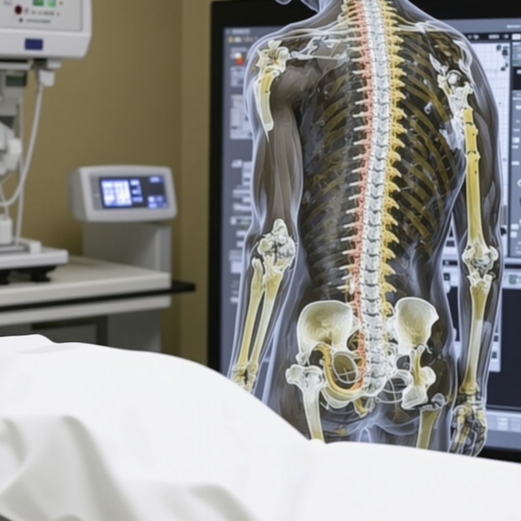
My Personal Encounter with Back Pain and the Search for Answers
Like many people, I’ve faced the frustration of persistent back pain that just wouldn’t go away. After trying various remedies, I finally decided to consult a specialist who recommended imaging tests to uncover the root cause. That’s when I started my journey into understanding the differences between MRI and X-ray for back pain diagnosis.
Unveiling the Secrets: What Makes MRI and X-ray Different?
In my research and experience, I found that X-rays are often the first step because they are quick, affordable, and effective at revealing bone-related issues. Conversely, MRI scans provide a detailed view of soft tissues, discs, nerves, and spinal cord. This distinction was crucial when my doctor explained that sometimes, X-rays might miss certain soft tissue injuries that only MRI can detect.
When Should You Opt for an MRI Instead of an X-ray?
During my consultation, I learned that if initial X-rays don’t reveal the cause of back pain, or if the pain persists despite treatment, an MRI might be necessary. For example, MRI is superior for diagnosing herniated discs, spinal stenosis, or nerve compression. According to a reputable source from the Spine-Health, MRI scans are invaluable for detailed soft tissue evaluation, which is often essential for comprehensive back pain management.
My Experience: Which Imaging Technique Gave Me the Clear Picture?
In my case, the MRI uncovered a herniated disc pressing on a nerve, which was not visible on the X-ray. That discovery changed my treatment plan entirely, leading to targeted therapies that finally alleviated my pain. It made me realize how important choosing the right imaging technique is for accurate diagnosis and effective treatment.
Is an MRI Always Better for Back Pain?
Not necessarily. X-rays are excellent for quick assessments of bone fractures or degenerative changes. However, for soft tissue injuries or nerve issues, MRI is the gold standard. It’s essential to consult with your orthopedic specialist to determine which imaging method aligns with your symptoms and medical history.
If you’re navigating back pain and unsure whether to pursue MRI or X-ray, I recommend discussing your options thoroughly with your healthcare provider. And if you’ve had similar experiences, I’d love to hear your story in the comments below! Sharing our journeys can help others make informed decisions about their health.
Understanding the Nuances of Back Pain Imaging: A Deep Dive
As an orthopedic specialist, I often encounter patients confused about which imaging technique is best suited for their specific back pain. While X-rays and MRIs are both invaluable tools, understanding their unique roles can significantly impact diagnosis accuracy and treatment outcomes. For instance, X-rays excel at revealing bony structures, fractures, or degenerative changes, making them a quick and cost-effective initial assessment. Conversely, MRI scans provide unparalleled detail of soft tissues, including discs, nerves, and the spinal cord, essential for diagnosing herniated discs or nerve compression.
Beyond the Basics: When Does MRI Become the Diagnostic Star?
One common question I hear is: “Is an MRI always necessary for back pain?” The answer hinges on the clinical scenario. If a patient presents with persistent pain unresponsive to conservative treatment, or if neurological symptoms such as numbness, weakness, or loss of bladder control are evident, an MRI is often indicated. This imaging modality can reveal subtle soft tissue injuries that X-rays might miss, guiding more targeted interventions. According to a comprehensive guide from the Spine-Health, MRI scans are particularly vital for evaluating disc herniations, spinal stenosis, and nerve impingements, especially when surgical intervention is being considered.
Case Study: My Personal Journey with MRI and X-ray
In my practice, I’ve seen cases where initial X-rays suggested degenerative changes but failed to explain persistent symptoms. An MRI later uncovered a herniated disc pressing on a nerve root, which was the true culprit. This discovery allowed for a precise, minimally invasive treatment plan, drastically improving the patient’s quality of life. It underscores the importance of selecting the appropriate imaging based on a thorough clinical evaluation. For more insights into non-surgical options, visit effective non-surgical care for herniated discs.
Expert Tip: Tailoring Imaging to Patient Needs
Choosing between MRI and X-ray isn’t always straightforward. Factors such as patient age, history, physical examination findings, and initial imaging results all influence the decision. For example, in cases of suspected spinal fractures, an X-ray might suffice initially, but if soft tissue injury is suspected, an MRI becomes indispensable. It’s crucial to collaborate closely with your orthopedic specialist to determine the most suitable imaging technique, ensuring an accurate diagnosis and effective treatment plan. Want to learn more about selecting the right imaging? Check out orthopedic support bracing options and latest innovations.
How Can I Ensure My Imaging Results Are Accurate and Useful?
Ensuring accurate imaging interpretation requires expert radiological review and correlating findings with clinical symptoms. Misinterpretation can lead to unnecessary treatments or missed diagnoses. Therefore, always seek care from experienced orthopedic radiologists and specialists who understand the nuances of spinal imaging. If you’re involved in a legal case or need documentation for insurance purposes, proper orthopedic documentation can make a significant difference, as discussed in orthopedic medical records for law firms.
If you’re navigating back pain and trying to decide whether MRI or X-ray is right for you, consult with a trusted orthopedic specialist. And if you’ve experienced similar diagnostic journeys, share your story in the comments below. Your insights can help others make informed health decisions!
My Journey into the Depths of Back Pain Imaging
As a seasoned orthopedic specialist, I often reflect on how crucial choosing the right imaging technique is. My personal experience began with a persistent back pain that stubbornly resisted initial treatments. I vividly remember the moment I realized that understanding the subtle differences between MRI and X-ray could make all the difference in diagnosis and treatment. This realization was a turning point in my approach to patient care and deepened my appreciation for the complexities involved in spinal imaging.
Unraveling the Layers: Why MRI Often Reveals What X-ray Misses
While X-rays are invaluable for quick assessments of bone integrity, they are limited in visualizing soft tissues. MRI, on the other hand, offers a detailed view of discs, nerves, and the spinal cord. I recall a case where an initial X-ray suggested degenerative changes, yet the real issue was a herniated disc pressing on a nerve root, only visible through MRI. This experience cemented my belief that MRI is often the more comprehensive tool when soft tissue injury is suspected, especially in cases where symptoms persist despite normal X-ray findings.
Deepening the Diagnostic Puzzle: When Is MRI Truly Essential?
One of the most sophisticated questions I encounter is: “How do I determine when an MRI is necessary beyond the initial X-ray?” The answer lies in clinical judgment. Persistent pain unresponsive to conservative treatments, neurological symptoms like numbness or weakness, or signs of nerve compression are strong indicators. According to authoritative sources like Spine-Health, MRI’s ability to reveal subtle soft tissue injuries often guides the decision for surgical intervention and tailored therapies. My own practice aligns with this, emphasizing a patient-centered approach that considers these nuanced factors.
Personal Reflection: The Power of Tailored Imaging in My Practice
Through years of experience, I’ve learned that personalized imaging strategies lead to better outcomes. For instance, I once managed a patient with chronic back pain where X-ray showed only mild degeneration. An MRI later revealed nerve impingement from a herniated disc, leading to targeted non-surgical treatment that transformed their quality of life. This reinforced my conviction that selecting the most appropriate imaging isn’t just a technical choice—it’s a profound decision that shapes patient journeys. If you’re curious about innovative, minimally invasive back pain treatments, I recommend exploring minimally-invasive back pain therapies.
Addressing the Broader Question: Is MRI Always Superior?
Not always. X-rays are quick, cost-effective, and excellent for detecting fractures or degenerative changes. MRI’s strength lies in soft tissue visualization. As I advise my patients, the choice depends on their specific symptoms and initial findings. Sometimes, a combined approach yields the best diagnostic clarity. For those navigating the maze of back pain diagnosis, I encourage open dialogue with your healthcare provider to ensure an imaging plan tailored to your needs. If you’ve had a similar diagnostic journey, sharing your story could help others understand their options better.
The Subtle Art of Advanced Imaging Decision-Making
In my practice, I emphasize the importance of integrating clinical findings with imaging results. For example, neurological deficits often necessitate MRI to assess nerve involvement accurately. It’s a nuanced process—balancing cost, urgency, and diagnostic yield. For those interested in the latest advancements, I recommend reviewing orthopedic support innovations that complement imaging to provide holistic care. Remember, the goal isn’t just imaging for its own sake but for crafting the most effective treatment strategy.
How Can Patients Advocate for the Right Imaging Tests?
Empowering yourself with knowledge is vital. Ask your doctor about the specific reasons for choosing MRI over X-ray, and how each will influence your treatment plan. Understanding the strengths and limitations of each modality helps in making informed decisions. If you’re involved in legal or insurance cases, accurate documentation and expert opinions can be pivotal, as discussed in orthopedic medical records for legal cases. Sharing your experiences can also provide valuable insights for others facing similar dilemmas.
The Nuances of Soft Tissue Visualization in MRI
One of the critical advantages of MRI scans lies in their unparalleled ability to visualize soft tissues, which are often the elusive culprits behind persistent back pain. During my extensive practice, I’ve observed that soft tissue injuries—such as ligament tears, subtle disc protrusions, or nerve entrapments—are frequently missed on X-rays but become evident on MRI. This capability is especially vital in cases where patients present with neurological symptoms like numbness or weakness that initial X-rays fail to explain. The detailed imaging provided by MRI enables clinicians to formulate precise treatment strategies, often avoiding unnecessary surgical interventions.
Integrating Advanced Imaging with Multidisciplinary Care
In my experience, the most effective back pain management strategies incorporate a multidisciplinary approach, leveraging the strengths of various diagnostic tools. For example, combining MRI findings with physical therapy, pain management, and minimally invasive procedures can dramatically improve outcomes. I often recommend patients explore minimally-invasive back pain treatments that are tailored based on comprehensive imaging results. This holistic approach ensures that treatment is not only targeted but also sustainable, reducing the likelihood of recurrence and chronicity.
Why Precision in Imaging Interpretation Matters for Legal and Insurance Cases
Accurate interpretation of MRI and X-ray results is paramount, especially in legal contexts where medical evidence can influence case outcomes. Misinterpretations or overlooked soft tissue injuries can lead to inadequate compensation or prolonged legal battles. As I emphasize in my practice, working closely with experienced radiologists and maintaining meticulous documentation can significantly strengthen your legal case. For detailed insights into ensuring your imaging reports are comprehensive and admissible, I encourage you to review orthopedic medical records for law firms.
How to Make the Most of Your Diagnostic Imaging Experience
Maximizing the benefits of MRI or X-ray involves understanding your specific symptoms and communicating effectively with your healthcare provider. Preparation may include providing a detailed history, following imaging instructions, and asking about the rationale for each test. I’ve found that informed patients tend to achieve better diagnostic accuracy because they are engaged in their care journey. If you’re curious about the latest innovations in spinal imaging or want to explore personalized treatment options, don’t hesitate to reach out or consult with a specialist who can tailor the diagnostic process to your unique needs.
Things I Wish I Knew Earlier (or You Might Find Surprising)
The Hidden Power of Soft Tissue Visualization
One thing I didn’t fully appreciate at first is how crucial MRI scans are for revealing soft tissue injuries. I used to think X-rays were enough for most back issues, but discovering nerve entrapments or disc tears often requires MRI’s detailed imaging. It’s a game-changer in diagnosis.
The Timing Matters More Than You Think
I learned that not every back pain needs an immediate MRI. Sometimes, a simple X-ray can rule out fractures or severe degenerative changes. But if symptoms persist or neurological signs appear, an MRI becomes essential. Knowing this helped me avoid unnecessary tests and focus on what truly mattered.
The Cost and Speed Trade-off
Initially, I was surprised at how much quicker and cheaper X-rays are compared to MRIs. They’re usually the first step, providing quick insights. MRI scans take longer and are more expensive, but they offer a deeper understanding when needed. It’s all about choosing the right tool at the right time.
Personal Stories Make a Difference
Listening to others’ experiences with back imaging has been eye-opening. Many people tell me how MRI revealed issues that X-rays missed, leading to better treatment outcomes. These stories remind me that personalized care depends on selecting appropriate imaging techniques.
When in Doubt, Consult a Specialist
My biggest takeaway is the importance of expert advice. A knowledgeable orthopedic specialist can help determine whether an X-ray or MRI is best for your specific situation. Don’t hesitate to ask questions and advocate for your health.
Resources I’ve Come to Trust Over Time
- Spine-Health: This site provides comprehensive and reliable information about back pain and imaging. It’s helped me understand when MRI is necessary and why.
- American Academy of Orthopaedic Surgeons (AAOS): Their guidelines on imaging choices are evidence-based and trustworthy, making them a great resource for patients and doctors alike.
- National Institutes of Health (NIH): NIH research articles and patient guides have deepened my understanding of the medical science behind back imaging techniques.
Parting Thoughts from My Perspective
Choosing between MRI and X-ray for back pain isn’t always straightforward, but understanding their strengths and limitations can significantly impact your diagnosis and treatment. From personal experience, I know that the right imaging can uncover hidden issues and guide effective care. If you’re facing back pain and unsure which test to pursue, I encourage you to discuss your symptoms thoroughly with your healthcare provider. Remember, informed decisions lead to better outcomes. If this resonated with you, I’d love to hear your thoughts or experiences—sharing our stories can help others navigate their own health journeys. Feel free to drop a comment below or explore more about minimally-invasive back pain treatments.

