Unlocking the Mystery Behind Orthopedic Imaging in New Jersey
In the realm of orthopedic care, precise diagnosis is the cornerstone of effective treatment. For residents of New Jersey facing musculoskeletal issues, understanding the spectrum of diagnostic and imaging services available is crucial. These advanced tools not only illuminate the root causes of pain but also guide orthopedic specialists toward tailored, minimally invasive interventions, enhancing recovery outcomes.
Beyond X-Rays: Exploring Cutting-Edge Orthopedic Imaging Modalities
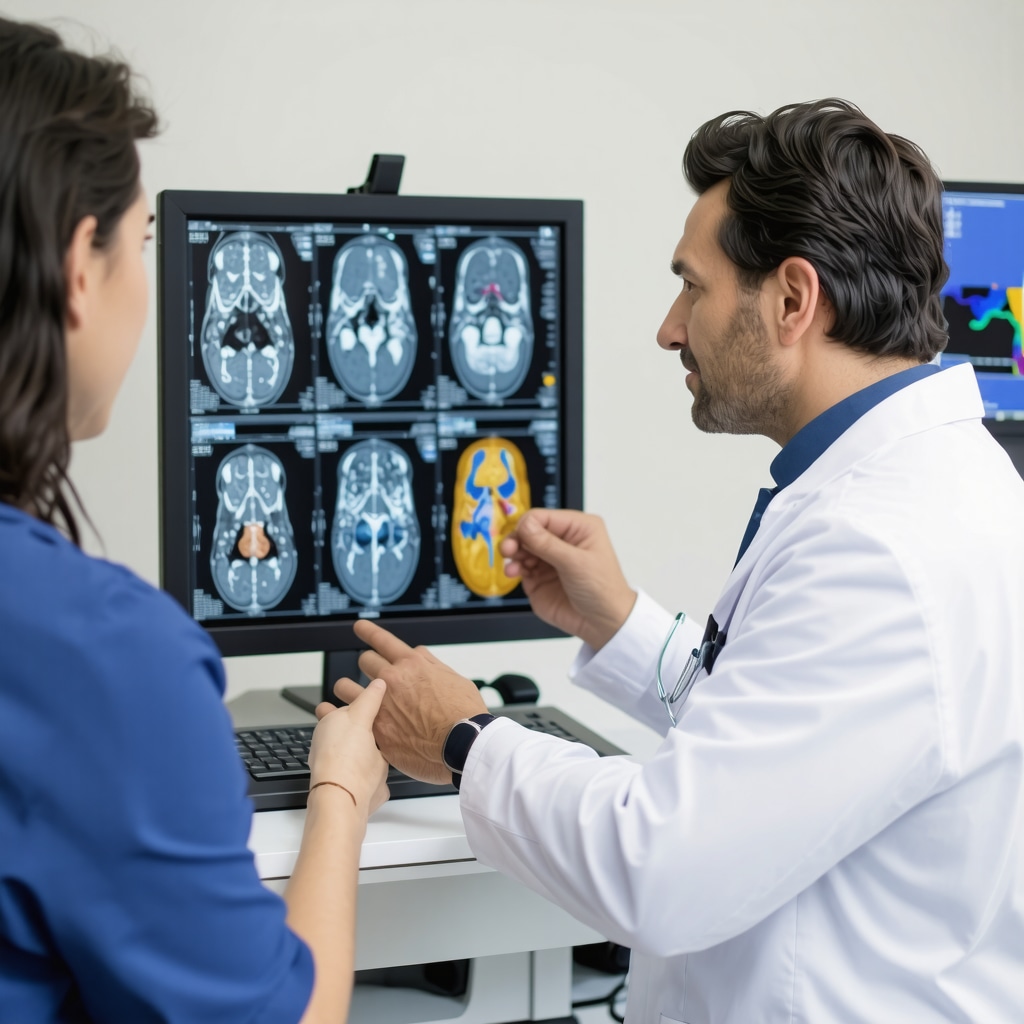
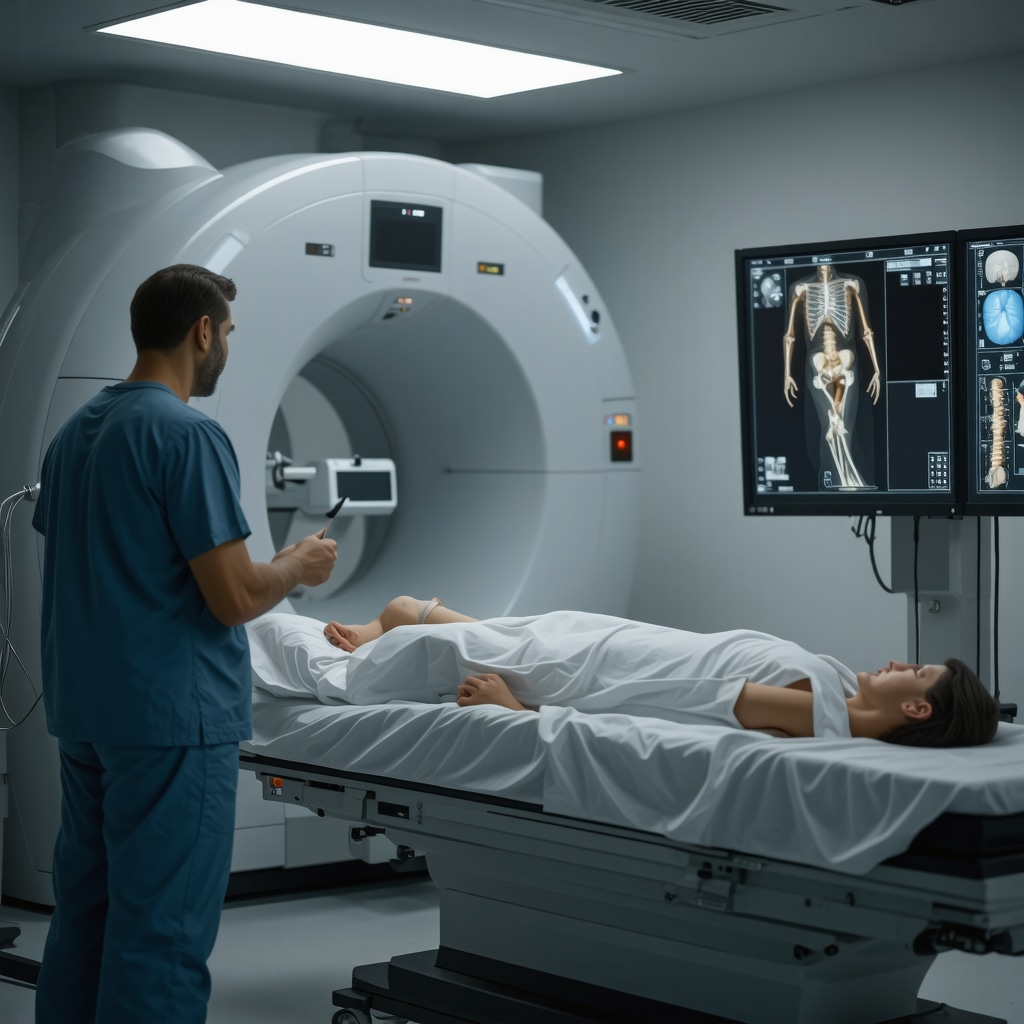
While X-rays remain a fundamental diagnostic tool, orthopedic imaging in NJ has evolved to encompass a myriad of sophisticated techniques. Magnetic Resonance Imaging (MRI) offers unparalleled soft tissue visualization, critical for diagnosing ligament tears and cartilage damage. Computed Tomography (CT) scans provide detailed cross-sectional images, essential in complex fracture assessments. Ultrasound imaging, increasingly popular in NJ clinics, delivers dynamic, real-time evaluation of tendons and muscles without radiation exposure. This multimodal approach allows clinicians to capture comprehensive insights that standard imaging alone cannot reveal.
How Do Orthopedic Specialists Decide Which Imaging Service Is Right for You?
Orthopedic experts in New Jersey weigh several factors when prescribing diagnostic imaging. Patient history, symptom severity, and the suspected injury type shape the decision-making process. For instance, a suspected rotator cuff tear might first be evaluated with an ultrasound, given its efficacy and accessibility. However, ambiguous symptoms or complex joint issues often necessitate MRI or CT scans for conclusive diagnosis. This tailored approach underscores the expertise involved in selecting the most informative and least invasive imaging method, ensuring patients receive precise care without unnecessary procedures.
Integrating Diagnostic Imaging with Advanced Orthopedic Care in NJ
The seamless integration of diagnostic imaging with therapeutic strategies is a hallmark of expert orthopedic care in New Jersey. Imaging guides not only initial diagnosis but also monitors treatment progress, from non-surgical interventions to post-operative rehabilitation. For example, patients undergoing minimally invasive back pain treatments benefit from imaging assessments that verify procedural success and inform rehabilitation protocols. This dynamic feedback loop exemplifies how imaging services empower both patient and clinician, fostering informed decisions and optimized recovery trajectories.
Experience Matters: Real-World Impact of Advanced Imaging in Orthopedic Outcomes
Consider the case of a New Jersey athlete experiencing persistent knee pain. Initial X-rays appeared normal, but an MRI revealed a subtle meniscal tear. Guided by this precise diagnosis, the orthopedic team implemented a targeted non-surgical regimen, sparing the patient from unnecessary surgery and enabling a swift return to sport. Such examples highlight the tangible benefits of advanced imaging in enhancing patient quality of life and streamlining care pathways.
For further expert insights on non-surgical orthopedic care, explore our detailed guide on effective non-surgical care for herniated discs.
Ready to Take the Next Step? Engage with NJ’s Orthopedic Imaging Experts
If precision and personalized care matter to you, connecting with NJ orthopedic specialists who leverage state-of-the-art diagnostic imaging can transform your treatment journey. Share your experiences or questions below — your insights might help others navigating similar challenges.
For authoritative information on musculoskeletal imaging, the Radiological Society of North America (RSNA) offers comprehensive resources that delve into the latest imaging technologies and their clinical applications. Visit their site for an in-depth exploration: Radiological Society of North America.
When Imaging Meets Experience: Navigating the Complexity Together
One thing I’ve come to appreciate over the years is how much trust plays a role in orthopedic imaging decisions. When I first faced recurring shoulder pain, the array of options—X-rays, MRIs, ultrasounds—felt overwhelming. It wasn’t just about the technology but finding a specialist who carefully listened and recommended the best path forward. This experience taught me that the most advanced imaging technology is only as good as the expertise interpreting it and the relationship built with the patient. In New Jersey, many orthopedic centers emphasize this balance, ensuring that technology complements strong clinical judgment.
Personalizing Imaging: Why One Size Doesn’t Fit All
Reflecting on my knee injury, I remember the initial X-rays showed no apparent damage, but pain persisted. The orthopedic team recommended an MRI, which uncovered a meniscal tear that would have otherwise gone unnoticed. This personalized approach made all the difference. It’s reassuring to know that imaging strategies can be tailored to your unique symptoms and lifestyle, rather than following a rigid protocol. This flexibility is especially important for athletes or active individuals in NJ who need precise diagnoses to avoid long-term issues.
How Can You Advocate for the Right Imaging to Get the Best Care?
It’s natural to wonder how you can be proactive when your doctor recommends imaging. From my experience, asking clear questions about why a specific imaging test is necessary and how it will influence your treatment can empower you. For example, a CT scan might be essential for complex fractures, but not always the first step. Understanding the reasoning helps you feel confident in your care plan.
Additionally, exploring resources from trusted organizations like the Radiological Society of North America can deepen your knowledge about imaging options. Being informed equips you to have meaningful discussions with your orthopedic specialist, ensuring your diagnostic journey is collaborative and transparent.
Integrating Imaging with Recovery: A Holistic Perspective
Post-diagnosis, imaging doesn’t just stop—it becomes a critical part of monitoring recovery. After my minimally invasive back treatment, follow-up imaging helped track healing progress and adjust rehabilitation exercises accordingly. This dynamic approach, common among NJ orthopedic professionals, ensures that treatment adapts to how your body responds, minimizing setbacks and promoting faster recovery.
If you’re curious about recovery support, check out our tips on orthopedic rehabilitation after lumbar fusion surgery. These insights emphasize how imaging and rehab go hand in hand for optimal outcomes.
Sharing Your Story: Community Wisdom in Orthopedic Care
Have you ever faced uncertainty about which imaging test to pursue or how to interpret the results? Your experience can offer valuable perspective to others navigating similar challenges. Feel free to share your journey or ask questions in the comments below. Engaging with others builds a supportive network, and sometimes, learning from real stories can be as powerful as clinical advice.
For those seeking expert orthopedic care in New Jersey, exploring options like top orthopedic spine specialists ensures you’re in skilled hands that prioritize both advanced imaging and individualized care.

Decoding Complex Orthopedic Cases: The Role of Functional and Quantitative Imaging Techniques
Beyond traditional imaging, New Jersey orthopedic specialists increasingly harness functional and quantitative imaging methods to unravel complicated musculoskeletal disorders. Techniques such as Dynamic Contrast-Enhanced MRI (DCE-MRI) and quantitative CT provide not only structural but also physiological and biomechanical data. This integration allows clinicians to assess tissue perfusion, inflammation, and bone mineral density with high precision, facilitating early detection of subtle pathologies like osteonecrosis or stress fractures that conventional imaging might miss.
For example, DCE-MRI can quantify synovial inflammation in rheumatoid arthritis patients, guiding targeted therapeutic interventions and monitoring response over time. Similarly, quantitative CT helps evaluate bone quality in osteoporosis, enabling preventive strategies before fractures occur. These advanced modalities empower orthopedic teams to tailor treatment plans based on comprehensive, multidimensional insights rather than solely anatomical abnormalities.
What Are the Latest Advances in Orthopedic Imaging That Improve Surgical Planning?
Recent innovations such as 3D imaging reconstruction and image-guided navigation have revolutionized surgical planning in orthopedics. Preoperative 3D models created from high-resolution CT or MRI scans allow surgeons to visualize a patient’s unique anatomy in exquisite detail, facilitating precise implant sizing and optimal surgical approach selection. Intraoperative imaging combined with navigation systems further enhances accuracy by providing real-time feedback, reducing risks of malalignment or incomplete repairs.
These technologies have been particularly transformative in complex spine surgeries and joint replacements, where millimeter-level precision significantly impacts functional outcomes and longevity of implants. Moreover, integration with robotic-assisted systems is paving the way for minimally invasive procedures with faster recovery and reduced complications.
Harnessing Artificial Intelligence: The Future of Orthopedic Imaging in New Jersey
Artificial intelligence (AI) and machine learning algorithms are rapidly emerging as powerful adjuncts in orthopedic imaging analysis. These tools can automatically detect subtle fractures, classify cartilage lesions, and quantify morphological changes with remarkable speed and consistency, surpassing traditional manual interpretation in some contexts.
In New Jersey’s leading orthopedic centers, AI-driven software assists radiologists by flagging potential abnormalities that may warrant further review, thus enhancing diagnostic sensitivity and reducing human error. Beyond detection, AI models are being developed to predict patient-specific outcomes based on imaging and clinical data, guiding personalized therapeutic decisions and rehabilitation protocols.
As these technologies mature, they promise to transform the diagnostic landscape by augmenting clinical expertise with data-driven insights, ultimately improving patient care quality and efficiency.
Integrating Imaging Data with Personalized Orthopedic Rehabilitation Programs
Modern orthopedic care in New Jersey increasingly emphasizes the synergy between imaging findings and individualized rehabilitation. Advanced imaging provides objective metrics to map injury extent and healing progression, which can be translated into customized physical therapy regimens.
For instance, quantitative MRI assessments of muscle atrophy or fatty infiltration post-injury can inform targeted strength training, while serial ultrasound evaluations of tendon healing guide graduated loading protocols. This precision rehabilitation approach minimizes the risk of re-injury and accelerates return to activity, especially for athletes and physically active patients.
Collaborative care teams, including orthopedic surgeons, radiologists, and physical therapists, utilize integrated imaging data to continuously adapt treatment plans, ensuring alignment with patient recovery milestones and functional goals.
How Does Advanced Imaging Influence Decision-Making in Complex Orthopedic Rehabilitation?
Advanced imaging informs not only the initial diagnosis but also nuanced decisions throughout rehabilitation. For example, persistent pain despite clinical improvement may prompt repeat MRI to evaluate for complications like scar tissue formation or incomplete healing. In cases of chronic tendinopathy, ultrasound elastography can assess tissue stiffness, guiding whether conservative management should continue or escalate to surgical intervention.
These insights enable clinicians to move beyond symptom-based treatment adjustments, adopting an evidence-driven approach that optimizes timing and modality of rehabilitation interventions. Consequently, patients benefit from more predictable recoveries and reduced incidence of chronic disability.
For a deeper dive into imaging-guided rehabilitation strategies, consult the comprehensive resources provided by the American Academy of Orthopaedic Surgeons (AAOS): AAOS Educational Resources.
Empowering Patients: How to Collaborate Effectively with Your Orthopedic Imaging Team
Active patient engagement is critical to maximizing the benefits of advanced orthopedic imaging. Understanding the purpose, scope, and implications of recommended imaging tests fosters shared decision-making and adherence to treatment plans.
Patients are encouraged to prepare thoughtful questions prior to imaging appointments, such as the expected diagnostic yield, potential risks, and how results will influence care pathways. Additionally, requesting visual explanations of imaging findings can enhance comprehension and trust.
New Jersey’s orthopedic practices often provide multidisciplinary consultations where radiologists and orthopedic surgeons jointly discuss imaging outcomes with patients, ensuring clarity and personalized guidance. This collaborative model exemplifies patient-centered care, empowering individuals to take an active role in their musculoskeletal health journey.
Revolutionizing Orthopedic Diagnostics: The Cutting-Edge of AI Integration
In the evolving landscape of orthopedic imaging, the infusion of artificial intelligence (AI) represents a paradigm shift for New Jersey specialists. AI algorithms now transcend traditional image interpretation by enabling automated detection of minute fractures, early cartilage degradation, and subtle morphological changes that often elude even seasoned radiologists. This technological augmentation not only accelerates diagnostic workflows but also enhances accuracy, fostering timely and targeted interventions.
How Is AI Shaping Personalized Treatment Plans in Orthopedic Imaging?
AI-driven analytics synthesize vast imaging datasets alongside patient-specific clinical parameters, generating predictive models that forecast disease progression and response to therapies. For instance, machine learning tools can stratify patients with osteoarthritis based on cartilage wear patterns visualized on MRIs, informing customized therapeutic regimens ranging from conservative management to surgical options. Furthermore, AI-assisted real-time intraoperative imaging guidance minimizes surgical errors, optimizing implant positioning and preserving native tissue integrity.
Quantitative Imaging Metrics: Unlocking Biomechanical Insights for Tailored Rehabilitation
Beyond morphological assessment, quantitative imaging modalities like T2 mapping and diffusion tensor imaging (DTI) provide granular insights into tissue microstructure and biomechanical properties. These metrics enable clinicians to objectively measure muscle quality, tendon elasticity, and cartilage composition, thereby refining rehabilitation protocols to individual biomechanical deficits. The integration of such imaging biomarkers into rehabilitation frameworks ensures progressive loading strategies that mitigate reinjury risk while expediting functional recovery.
Collaborative Multidisciplinary Approaches: Bridging Imaging and Clinical Expertise
The confluence of radiologists, orthopedic surgeons, and rehabilitation specialists in New Jersey fosters an interdisciplinary environment where imaging findings translate directly into clinical decision-making. Multimodal imaging reviews coupled with biomechanical assessments empower teams to construct holistic, patient-centered management plans. This synergy enhances the precision of interventions and aligns with contemporary value-based care models emphasizing outcomes and patient satisfaction.
For clinicians and patients seeking to deepen their understanding of advanced imaging applications in orthopedics, the American College of Radiology (ACR) provides authoritative guidelines and case studies elucidating best practices: American College of Radiology Clinical Resources.
Taking the Next Step: Engage with New Jersey’s Pioneers in Orthopedic Imaging
Are you ready to leverage the transformative power of AI-enhanced and quantitative imaging to elevate your orthopedic care? Connect with New Jersey’s leading experts who seamlessly integrate these innovations into personalized treatment pathways. Share your questions or experiences below to join a community dedicated to advancing musculoskeletal health through cutting-edge diagnostics.
Frequently Asked Questions (FAQ)
What types of orthopedic imaging are most commonly used in New Jersey, and how do they differ?
Orthopedic imaging in New Jersey commonly includes X-rays, MRI, CT scans, and ultrasound. X-rays excel at visualizing bone fractures and alignment, MRI provides superior soft tissue contrast for ligaments, cartilage, and muscles, CT offers detailed cross-sectional images useful for complex fractures, and ultrasound supplies real-time dynamic assessment of tendons and muscles without radiation exposure. Each modality is selected based on the suspected pathology and clinical context.
How do orthopedic specialists decide which imaging modality to recommend for a patient?
Specialists consider factors such as patient history, symptom severity, suspected injury type, and previous imaging results. For example, an ultrasound may be preferred initially for suspected tendon injuries due to accessibility and safety, while MRI or CT is chosen for complex joint or bone evaluations. The goal is to obtain precise diagnostic information with minimal invasiveness and radiation exposure.
What role does artificial intelligence play in modern orthopedic imaging?
AI enhances image interpretation by detecting subtle abnormalities like microfractures or early cartilage degeneration with high accuracy and speed. It assists radiologists by flagging areas of concern, supports predictive modeling for disease progression, and guides personalized treatment planning. Intraoperative AI integration also improves surgical precision and outcomes.
Can advanced imaging techniques improve rehabilitation outcomes?
Absolutely. Quantitative imaging methods, such as T2 mapping and ultrasound elastography, provide objective data on tissue quality and healing status. This information enables clinicians to tailor rehabilitation protocols to individual biomechanical needs, optimizing strength training and reducing re-injury risk. Continuous imaging feedback helps adjust therapies dynamically throughout recovery.
Are there risks associated with orthopedic imaging?
Most orthopedic imaging modalities are safe; however, X-rays and CT scans involve ionizing radiation, so their use is judiciously balanced against diagnostic benefit. MRI and ultrasound do not use radiation and are generally safe for most patients. Patients should discuss any concerns or contraindications, such as metal implants or pregnancy, with their providers.
How can patients actively participate in decisions about their imaging and care?
Patients are encouraged to ask why a particular imaging test is recommended, what information it provides, and how it will affect treatment options. Reviewing imaging results with specialists and seeking visual explanations fosters understanding and trust. Utilizing resources from reputable organizations like the Radiological Society of North America enhances patient empowerment.
What advancements in imaging technology are shaping the future of orthopedic diagnostics in New Jersey?
Emerging trends include 3D imaging reconstruction, image-guided navigation, robotic-assisted surgery, and AI-driven analytics. These innovations allow for unprecedented anatomical visualization, enhanced surgical accuracy, and personalized treatment strategies, ultimately improving patient outcomes and reducing recovery times.
How does multidisciplinary collaboration enhance orthopedic imaging effectiveness?
Collaboration among radiologists, orthopedic surgeons, and rehabilitation specialists ensures imaging findings are integrated with clinical and biomechanical assessments. This team approach facilitates comprehensive, patient-centered care plans that adapt to evolving recovery stages and functional goals, promoting better long-term results.
Is specialized orthopedic imaging available for athletes in New Jersey?
Yes, many centers offer tailored imaging services designed for athletes, focusing on early detection of overuse injuries, cartilage wear, and soft tissue damage. Dynamic and quantitative imaging techniques help guide safe return-to-play decisions and prevent chronic conditions.
How do imaging techniques help in managing chronic orthopedic conditions like arthritis?
Advanced imaging modalities such as Dynamic Contrast-Enhanced MRI can quantify inflammation and tissue changes, enabling precise monitoring of disease activity and response to therapy. Quantitative CT assesses bone density, guiding preventive measures for osteoporosis-related fractures. These insights support personalized treatment adjustments and improved quality of life.
Trusted External Sources
- Radiological Society of North America (RSNA): Provides in-depth resources on musculoskeletal imaging technologies and clinical applications, ensuring updated knowledge on diagnostic advancements.
- American Academy of Orthopaedic Surgeons (AAOS): Offers educational materials on imaging-guided orthopedic care and rehabilitation protocols, bridging clinical practice with imaging insights.
- American College of Radiology (ACR): Publishes authoritative guidelines and case studies on best practices in orthopedic imaging, fostering multidisciplinary collaboration and quality standards.
- National Institute of Arthritis and Musculoskeletal and Skin Diseases (NIAMS): Supplies research-based information on musculoskeletal disorders and the role of imaging in diagnosis and management.
- Journal of Orthopaedic Research: Features peer-reviewed studies on novel imaging techniques and their clinical impact, supporting evidence-based orthopedic practice.
Conclusion
Orthopedic imaging in New Jersey has transcended traditional boundaries through the integration of advanced modalities, artificial intelligence, and collaborative clinical expertise. From precise diagnostics to personalized rehabilitation strategies, these innovations empower patients and clinicians alike to navigate complex musculoskeletal conditions with confidence. Understanding the nuances of imaging options, actively engaging with your care team, and leveraging trusted resources can significantly enhance treatment outcomes. If you or a loved one face orthopedic challenges, consider consulting with New Jersey’s leading specialists who blend cutting-edge imaging technology with individualized care. Share your experiences or questions below, and explore more expert content to stay informed and proactive in your orthopedic health journey.


Having recently gone through a knee injury, I can personally attest to the importance of advanced imaging techniques like MRI in uncovering issues that X-rays might miss. The post’s explanation of how orthopedic specialists in New Jersey customize imaging choices based on individual symptoms really resonated with me. It’s reassuring to know that ultrasounds can be a radiation-free first step for tendon evaluation, which is often overlooked. One aspect I found particularly insightful was the integration of imaging with ongoing treatment—especially how it guides rehabilitation by tracking healing progress. This dynamic approach helps avoid setbacks and tailors therapy to actual recovery status rather than just symptoms. I’m curious how others have advocated for specific imaging tests during their consultations. Have you ever felt the need to question or request a particular type of scan to ensure your diagnosis was accurate? What strategies have worked for you in communicating effectively with your orthopedic team to get the best personalized care? I’d love to hear about experiences or tips from fellow readers navigating these diagnostic decisions in NJ.
Replying to Emily Richards’ insightful comment — I completely agree that advocating for the right imaging test is crucial. In my experience with a shoulder injury, I initially received an X-ray that showed no issues, but my pain persisted. It wasn’t until I asked my orthopedic specialist about getting an MRI that we uncovered a partial rotator cuff tear. What really helped me communicate effectively was coming prepared with specific questions about each imaging option’s benefits and limitations, much like the post suggests. Also, I found that requesting to see the images during consultations and having the radiologist explain the findings made a big difference in understanding my condition and treatment plan. I’m curious, has anyone else found that multidisciplinary discussions involving radiologists improve their confidence in the imaging results? It seems that New Jersey practices emphasizing this collaborative approach truly enhance patient trust and outcomes. Moreover, integrating imaging with the rehab process has been a revelation in my recovery — knowing exactly how my tissue was healing informed my physical therapist’s adjustments. How do others feel about the balance between technology and clinical judgment in managing musculoskeletal injuries? It seems that no single modality or test fits all, and the personalized approach detailed here is the way forward.
Reading about how orthopedic imaging has evolved beyond just X-rays in New Jersey truly highlights the importance of personalized care. I especially appreciate the focus on how specialists select imaging based on individual symptoms, history, and injury type rather than a one-size-fits-all approach. From my own experience, opting for an ultrasound first for a suspected tendon injury was a great step—quick, radiation-free, and insightful for my condition. What stood out to me in the post is the seamless integration of imaging from diagnosis through treatment and rehab, helping adjust care as healing progresses. This dynamic monitoring gives patients a better sense of involvement and clarity about their recovery. I also find it promising that advanced techniques like functional MRI and AI-assisted imaging are becoming part of orthopedic care in NJ, offering more precise insights. Given these advances, I wonder how others perceive balancing reliance on cutting-edge technology with the essential clinical judgment and patient communication that specialists bring. How do you think patients can best advocate for imaging choices that truly fit their needs without feeling overwhelmed? I’m curious to hear others’ strategies or stories around collaborating closely with imaging teams during their orthopedic journey.
The post’s emphasis on personalized imaging approaches truly resonates with me, especially considering how every patient’s symptoms and lifestyles differ. I once faced a foot injury where initial X-rays didn’t reveal much, but the orthopedic team recommended an ultrasound to assess tendon involvement, which turned out to be crucial for my diagnosis and subsequent treatment. I appreciate how New Jersey orthopedic specialists balance technological advances with clinical experience — it’s not just about having the latest tools, but selecting the right one for each situation. Given how complex and sometimes overwhelming imaging options can be, I found that preparing questions beforehand and asking my specialist to explain how each test ties into my treatment helped me feel in control rather than overwhelmed. Also, the integration of imaging throughout recovery was essential, as it allowed adjustments to my rehab rather than relying only on symptom reports. I’m curious how others approach situations where multiple imaging options are available—do you prefer trusting your doctor’s judgment entirely, or do you research and advocate for certain modalities yourself? How do you ensure you get the imaging that truly benefits your care without unnecessary procedures? Sharing strategies could help many in this community better navigate their orthopedic journeys.
I really appreciate how this post highlights the crucial role of advanced imaging modalities in tailoring orthopedic care to individual patients in New Jersey. From my experience recovering from a rotator cuff issue, the choice between ultrasound and MRI can feel daunting. Ultrasound was my initial test — it’s great for real-time assessment and carries no radiation risk — but it was the MRI that uncovered the extent of my injury, providing detailed insight necessary for an effective treatment plan. One aspect that stood out to me is the integration of imaging throughout the rehabilitation process; seeing objective evidence of healing via follow-up scans really enhanced my confidence in the treatment and helped my therapists adjust exercises appropriately. I also find the emerging use of AI and quantitative imaging fascinating in predicting outcomes and customizing rehabilitation strategies. That said, I sometimes wonder how patients can balance trusting clinical judgment with the temptation to push for advanced imaging out of anxiety or uncertainty. How have others navigated discussions with their orthopedic team about balancing necessary imaging versus avoiding over-testing? For those who’ve used AI-informed imaging, have you noticed tangible benefits in your treatment outcomes? I’d love to hear different perspectives on this evolving landscape of orthopedic diagnostics in NJ.
I found the detailed explanation about how orthopedic specialists in New Jersey decide between imaging options especially insightful. It really highlights that there’s no universal solution when it comes to diagnostics – each patient’s history, symptoms, and injury complexity demand tailored imaging choices. From personal experience with a sports injury, I initially underwent X-rays which showed nothing definitive, but persistent discomfort led my doctor to recommend an MRI. That transition was crucial because the MRI unveiled a partial ligament tear that otherwise would have gone undetected.
What struck me most is the growing role of ultrasound as a first line, non-invasive tool, which is not only efficient but also spares patients from radiation exposure. However, navigating when to push for more advanced imaging like CT or MRI can be tricky for patients. It’s encouraging that New Jersey orthopedic centers often facilitate multidisciplinary discussions, where radiologists and orthopedic surgeons jointly review images and outcomes with patients. This kind of communication fosters trust and patient empowerment.
I’m curious how others feel about integrating modern AI-driven analysis into this process—have you experienced greater confidence or faster diagnoses with AI-assisted imaging? Also, how do you balance relying on clinical judgment versus advocating for additional imaging when uncertainties remain? I’d love to hear different approaches to staying informed but not overwhelmed in such decisions.