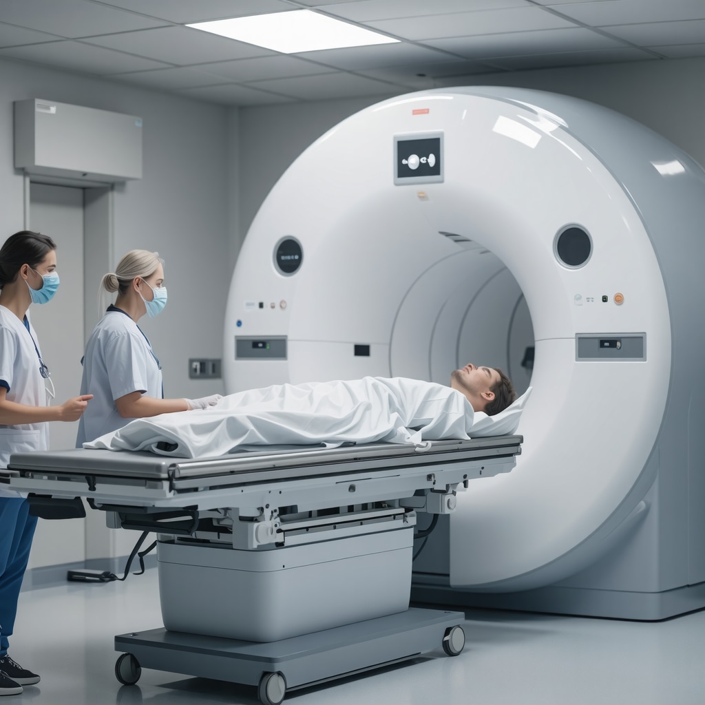
My Personal Journey with Orthopedic Diagnostic Imaging
Recently, I found myself in need of an MRI scan to evaluate a lingering shoulder issue. Like many, I was initially overwhelmed by the idea of medical imaging, unsure of what to expect or how to prepare. Sharing my experience, I hope to shed light on the process and ease any fears you might have about your upcoming scan.
Understanding the Importance of Proper Preparation
From my experience, preparing adequately for an orthopedic diagnostic imaging appointment can make a significant difference. It not only ensures clear, accurate images but also helps avoid unnecessary repeat scans. I learned that knowing what to do before your scan can streamline the process and reduce anxiety.
How I Got Ready for My MRI: Practical Tips and Insights
One of the first things I did was confirm whether I should fast or avoid certain medications. My doctor advised me to wear comfortable, loose-fitting clothing without metal fasteners, which I found helpful. I also removed all jewelry and metallic accessories to prevent interference with the MRI machine. These steps might seem simple, but they are crucial for a smooth experience.
What to Expect During Your Imaging Appointment
During my scan, I was surprised by how quiet and comfortable the environment was. The technician explained the procedure clearly and reassured me throughout. The entire process lasted about 30 minutes, and I was able to listen to music through headphones, which made the time pass quickly. Knowing what to expect really eased my nerves.
Why Accurate Imaging Matters for Your Orthopedic Care
Accurate diagnostic images are vital for effective treatment planning. For example, if you’re dealing with a herniated disc or ligament injury, a clear MRI or X-ray can reveal the extent of damage and guide your doctor in choosing the right treatment, whether surgical or non-surgical. I found that thorough imaging paved the way for targeted and successful recovery.
Have you ever wondered how different imaging techniques compare in diagnosing spinal or joint issues?
Research indicates that MRI scans provide detailed images of soft tissues, making them ideal for diagnosing ligament tears or disc herniations, while X-rays are better suited for evaluating bone fractures or alignment issues. Understanding these differences helped me appreciate the importance of choosing the appropriate imaging method for my condition.
If you’re preparing for an orthopedic diagnostic scan, I recommend discussing all your concerns with your healthcare provider. They can give you tailored instructions and support. And if you’re curious about other aspects of orthopedic care, check out rehab tips after lumbar fusion.
Feel free to share your experiences or questions in the comments below. Your insights might help others navigate their own diagnostic journeys more confidently!
Decoding the Complexities of Orthopedic Imaging Techniques for Spinal Health
As an orthopedic specialist, I often encounter patients overwhelmed by the variety of diagnostic imaging options available for spinal issues. Understanding when to opt for an MRI, CT scan, or X-ray is crucial in crafting an effective treatment plan. For instance, while X-rays excel at revealing bone alignment and fractures, MRI scans provide unparalleled detail of soft tissues, such as discs, ligaments, and nerves, making them invaluable for diagnosing herniated discs or ligament tears. A nuanced approach ensures that patients receive precise diagnoses without unnecessary procedures.
What Are the Practical Implications of Imaging Choices in Orthopedic Care?
Choosing the appropriate imaging modality directly impacts treatment strategies. An MRI revealing a disc herniation might lead to conservative management like physical therapy or targeted injections, whereas clear bone fractures from an X-ray could necessitate surgical intervention, such as spinal fusion. Moreover, from a legal standpoint, detailed and accurate imaging documentation can significantly influence personal injury claims—highlighting the importance of selecting the right diagnostic tool early in the process. To explore effective non-invasive treatments, visit this resource.
How Do Emerging Imaging Technologies Improve Diagnostic Precision?
Advancements like 3D imaging and functional MRI are revolutionizing orthopedic diagnostics. These technologies enable clinicians to assess not only static anatomical structures but also dynamic functions, such as nerve conduction or blood flow, providing a comprehensive picture of spinal health. Incorporating these innovations can lead to earlier detection of degenerative changes and more personalized treatment plans. For patients, this means less guesswork and quicker relief. If you’re considering imaging options, consult your healthcare provider about the latest advancements and how they might benefit your specific condition.
Have you considered how imaging choices influence long-term outcomes in spinal care?
In-depth knowledge of each modality’s strengths and limitations allows clinicians to tailor interventions effectively. For example, selecting an MRI over an X-ray for suspected nerve impingement ensures soft tissue damage is not overlooked, potentially avoiding unnecessary surgeries later. This strategic decision-making underscores the importance of expert consultation when navigating spinal diagnostics. If you’re unsure about which imaging is suitable for your situation, a visit to a specialized orthopedist can clarify your options. To find a trusted specialist near you, check this guide.
Engaging with your healthcare team about the benefits and limitations of each imaging technique can empower you to make informed decisions. Feel free to share your experiences or ask questions in the comments below—your insights might help others make better choices in their orthopedic journeys!
Unveiling the Depths of Diagnostic Precision in Orthopedics
Reflecting on my journey through orthopedic diagnostics, I realize that choosing the right imaging technique is akin to solving a complex puzzle where each piece offers unique insights. During my practice, I’ve observed how MRI scans, with their unparalleled soft tissue clarity, can reveal subtle nerve impingements that might be missed on traditional X-rays. This nuanced understanding underscores the importance of tailoring imaging choices not just to the obvious symptoms but also to the intricate details that can influence treatment outcomes.
The Subtle Art of Personalizing Imaging Strategies
One of the most fascinating aspects I’ve encountered is the evolving landscape of advanced imaging technologies like functional MRI and 3D imaging. These innovations allow us to visualize dynamic processes, such as blood flow or nerve conduction, providing a more holistic view of spinal health. Personally, I find that integrating these tools into diagnostic protocols demands both technical expertise and a keen eye for detail—skills honed over years of experience. This approach enables a more personalized treatment plan, aligning with each patient’s unique anatomy and pathology.
Deepening the Dialogue: How Do Imaging Choices Shape Long-Term Outcomes?
It’s intriguing to consider how initial imaging decisions can ripple through the entire treatment trajectory. For instance, selecting an MRI over an X-ray for suspected soft tissue injuries can prevent misdiagnosis, reduce unnecessary procedures, and ultimately enhance recovery. Studies, such as those published in the Journal of Orthopaedic Surgery & Research, emphasize that early, accurate diagnosis driven by appropriate imaging correlates strongly with better long-term results. This insight reinforces my commitment to meticulous imaging selection as a cornerstone of effective orthopedic care.
What Are the Ethical and Practical Considerations in Advanced Imaging?
While cutting-edge technologies offer remarkable benefits, they also introduce ethical dilemmas—such as cost-effectiveness and accessibility. Balancing patient benefit with resource allocation requires careful judgment. From a practical standpoint, I advocate for a thoughtful, evidence-based approach: leveraging advanced imaging when it genuinely alters management, rather than as a routine. Engaging patients in shared decision-making about these options fosters trust and ensures that technological advancements serve their best interests.
If you’ve experienced the complexities of orthopedic diagnostics firsthand or are curious about how to optimize your imaging choices, I invite you to share your insights or questions in the comments. Your stories can illuminate the nuanced realities faced by many on their path to recovery.
The Future of Orthopedic Imaging: Embracing Innovation and Personalization
Looking ahead, I am excited about the potential of artificial intelligence and machine learning to revolutionize diagnostic accuracy. These tools can analyze vast imaging datasets, identify patterns beyond human perception, and suggest personalized treatment pathways. As practitioners and patients, staying informed about these developments will be crucial in making educated choices and advocating for the most effective, least invasive diagnostics available. For those interested in exploring the latest innovations, visiting trusted sources like top spine specialists in 2025 can provide valuable insights.
Ultimately, the intersection of technology, expertise, and personalized care defines the future of orthopedic imaging—a journey I am privileged to be part of, continually learning and adapting to serve my patients better.
Deciphering the Nuances of Spinal Imaging for Precise Long-Term Outcomes
In my ongoing journey through orthopedic diagnostics, I’ve come to appreciate the profound impact that nuanced imaging choices have on patient prognosis. Advanced modalities like functional MRI and 3D imaging are not merely technological luxuries but essential tools that unveil the subtle complexities of spinal pathology. For example, while traditional MRI provides static snapshots, functional MRI can assess nerve conduction and blood flow, revealing early degenerative changes that might otherwise go unnoticed. These insights enable clinicians to tailor interventions with remarkable precision, ultimately enhancing long-term recovery and quality of life.
The Ethical and Practical Dimensions of Embracing Cutting-Edge Imaging
Integrating innovative imaging techniques raises critical ethical considerations, particularly regarding cost-effectiveness and equitable access. As a practitioner, I advocate for a judicious approach—reserving advanced diagnostics for cases where they significantly influence treatment decisions. This strategy aligns with evidence-based practices, ensuring optimal resource utilization without compromising patient care. Engaging patients in transparent discussions about the benefits, limitations, and costs of these technologies fosters trust and shared decision-making. For further insights on balancing innovation with practicality, explore this comprehensive guide.
Harnessing AI and Machine Learning to Elevate Diagnostic Precision
The future of orthopedic imaging is poised for transformative advancements through artificial intelligence and machine learning. These technologies can analyze vast datasets, identify subtle patterns, and predict disease progression with unprecedented accuracy. For instance, AI algorithms can differentiate between benign and malignant soft tissue anomalies, reducing diagnostic uncertainty. As clinicians and patients, staying informed about these developments enables us to advocate for smarter, less invasive diagnostics. If you’re interested in how these innovations are shaping personalized treatment plans, I encourage you to connect with leading specialists through top spine specialists in 2025.
Deepening the Dialogue: How Can We Optimize Imaging Strategies for Better Outcomes?
Optimizing imaging strategies requires a delicate balance between technological capability and clinical necessity. It involves continuous education, interdisciplinary collaboration, and mindful patient engagement. For example, selecting an MRI over an X-ray for suspected nerve impingement ensures soft tissue integrity is accurately assessed, guiding effective interventions that prevent long-term disability. Moreover, meticulous documentation and clear communication with patients about the rationale behind each imaging choice can significantly improve adherence to treatment plans. I invite you to share your experiences or questions about personalized imaging approaches in the comments—your insights can inspire others to make informed decisions.
The Evolving Landscape of Orthopedic Diagnostics: A Personal Reflection
Reflecting on my professional evolution, I recognize that embracing technological innovation while maintaining clinical judgment is the cornerstone of effective orthopedic care. The integration of advanced imaging techniques has expanded our diagnostic toolkit, enabling us to detect and address issues at their earliest stages. This proactive approach not only improves patient outcomes but also minimizes invasive procedures and accelerates recovery. As we navigate this dynamic landscape, continuous learning and adaptation are essential. For more about how cutting-edge diagnostics can revolutionize your care, visit this resource for comprehensive rehabilitation strategies.
Things I Wish I Knew Earlier (or You Might Find Surprising)
1. The Power of Soft Tissue Imaging
Early in my experience, I underestimated how crucial MRI scans are for soft tissue evaluation. I thought X-rays were sufficient for most issues, but discovering ligament tears and disc herniations required that detailed imaging opened my eyes to the importance of choosing the right test from the start.
2. Preparation Can Make or Break Your Scan
Simple steps like removing all jewelry and wearing loose clothing can significantly improve the quality of your images. I wish I had realized how much my preparation influences the accuracy of diagnostics and, ultimately, my treatment plan.
3. Not All Imaging Is Created Equal
Understanding that X-rays are best for bones, while MRIs excel at soft tissues, helped me advocate for myself. This knowledge empowered me to ask better questions and ensure my doctor ordered the most appropriate test for my condition.
4. Technology Is Rapidly Evolving
Advancements like 3D imaging and functional MRI are changing the game. I was amazed to learn how these innovations can reveal early degenerative changes and personalize treatment strategies, which I hadn’t appreciated before.
5. Cost and Accessibility Matter
While cutting-edge imaging offers detailed insights, it can be costly and less accessible. Striking a balance between technological benefits and practical considerations is something I’ve come to value deeply.
6. Trust Your Healthcare Team
Open communication with your doctors about the purpose and necessity of each imaging test builds trust and leads to better care. I’ve found that being informed makes the whole process less intimidating and more collaborative.

