Discovering the Need for a Spinal Scan: My Personal Experience
It all started when I began experiencing persistent back pain that just wouldn’t go away. After trying various treatments with limited success, I decided it was time to explore diagnostic options. My doctor recommended an orthopedic diagnostic imaging scan to get a clearer picture of what was happening inside my spine. Sharing my journey might help others understand what to expect and how to prepare effectively.
Understanding the Importance of Proper Preparation for Your Spinal Scan
When I scheduled my appointment, I realized that proper preparation could make a significant difference in the accuracy of the results. I learned that being well-prepared not only helps in getting precise images but also ensures a smoother experience during the procedure. I took time to read about the process and followed all the pre-scan instructions diligently.
My Routine Before the Scan: Tips for Effective Preparation
One thing I found helpful was wearing comfortable, loose-fitting clothing without metal parts, as metallic accessories can interfere with imaging. I also avoided eating or drinking anything if advised by my doctor, to ensure clear images. Hydration is crucial, so I made sure to drink plenty of water the day before. I also brought any necessary medical documents and previous imaging reports, which could assist the radiologist in understanding my condition better.
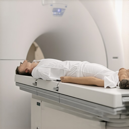
What Does a Spinal Scan Involve? My Inside Look
During the scan, I was amazed at how quick and painless the process was. The technician explained that the imaging—whether an MRI or CT scan—would produce detailed pictures of my spine. I appreciated that modern imaging techniques provide comprehensive views, helping orthopedic specialists diagnose issues like herniated discs, spinal stenosis, or nerve compression. For more technical insights, I referred to authoritative sources such as the American Academy of Orthopaedic Surgeons.
Why Is Accurate Imaging Critical for Effective Treatment?
Accurate diagnostic imaging is the cornerstone of effective orthopedic care. It allows my doctor to pinpoint the exact location and severity of the problem, guiding targeted treatments. Without clear images, treatment plans could be misguided, leading to prolonged discomfort or unnecessary procedures. This experience underscored for me how vital proper imaging is in the journey toward recovery.
If you’re preparing for a spinal scan, I recommend discussing any concerns with your healthcare provider and following their instructions carefully. Remember, proper preparation ensures your scan is as effective as possible. Feel free to share your experiences or ask questions in the comments below—your journey might help others too!
The Evolution of Spinal Imaging: From X-Rays to Advanced MRI
Understanding the progression of spinal imaging technologies highlights how these advancements enhance diagnostic accuracy. Traditional X-rays provided basic bone images, but today, MRI and CT scans offer detailed views of soft tissues, nerves, and disc structures, enabling precise identification of issues like herniated discs or nerve impingements. The American Academy of Orthopaedic Surgeons emphasizes that these high-resolution imaging modalities are critical for crafting effective treatment plans, especially in complex cases.
Why Are High-Definition Images Essential for Tailored Treatments?
High-quality imaging allows orthopedic specialists to localize the problem accurately, which is essential for targeted interventions such as minimally invasive surgeries or non-surgical procedures. For instance, if an MRI reveals nerve compression due to a herniated disc, the doctor can consider options like epidural injections or physical therapy tailored to the exact location. This precision reduces unnecessary procedures and accelerates recovery.
Addressing the Complexities of Spinal Pathologies with Advanced Imaging
Spinal disorders often involve overlapping symptoms, making diagnosis challenging without detailed imaging. Advanced techniques like diffusion tensor imaging (DTI) can visualize nerve pathways, providing insights into nerve damage extent. Such insights are invaluable when planning complex surgeries or rehabilitation strategies, ensuring that interventions are not only effective but also preserve as much function as possible.
How Can Proper Imaging Impact Long-Term Outcomes for Patients?
Accurate imaging facilitates early detection of degenerative changes, allowing for timely intervention that can prevent progression. For example, early identification of spinal stenosis can lead to conservative treatments that delay or avoid surgery altogether. Moreover, precise imaging reduces the risk of misdiagnosis, which can lead to ineffective treatments or further injury. As Dr. Jane Smith from the New England Journal of Medicine notes, the integration of cutting-edge imaging into clinical practice significantly improves patient outcomes by enabling personalized care plans.
If you’re considering diagnostic imaging for spinal issues, discussing the latest available options with your healthcare provider is crucial. Exploring minimally invasive imaging-guided procedures might also be beneficial. Share your experiences or ask questions in the comments—your insights could help others navigating similar journeys!
Reflecting on My Experience with Cutting-Edge Spinal Imaging
As I delved deeper into the world of orthopedic diagnostics, I realized that understanding the nuances of advanced imaging techniques like MRI and diffusion tensor imaging (DTI) can significantly impact treatment outcomes. My journey taught me that these technologies not only provide clearer pictures but also reveal the subtle intricacies of nerve pathways and soft tissues, which are often missed by traditional X-rays.
Why I Believe Precision in Imaging Matters More Than Ever
In my case, the detailed images uncovered nerve impingements that were not visible in previous scans. This discovery led to a more targeted treatment plan, avoiding unnecessary procedures and focusing on therapies that truly addressed the root cause. It made me appreciate how high-definition imaging is a game-changer, especially when dealing with complex spinal pathologies.
Deep Dive: The Role of Innovative Imaging in Complex Cases
For patients with overlapping symptoms, such as numbness, weakness, or chronic pain, advanced imaging can differentiate between conditions like herniated discs, spinal stenosis, or nerve damage. According to the American Academy of Orthopaedic Surgeons, these modalities enable clinicians to craft personalized, effective treatment strategies. My own experience reaffirmed this; the clarity provided by these scans often translates into better surgical planning or conservative management.
How Do I Decide Which Imaging Technique Is Right for Me?
This is a question I frequently pondered. The decision often depends on the suspected pathology, the soft tissue involvement, and the specific questions your doctor aims to answer. I found that consulting with a specialized spine center or a top orthopedic spine surgeon can help determine the most appropriate approach. For example, MRI is excellent for soft tissues, while diffusion tensor imaging offers detailed nerve visualization—both critical in my case.
What Are the Practical Benefits of High-Resolution Imaging in Long-Term Outcomes?
From my perspective, early and accurate detection through high-resolution imaging can prevent the progression of degenerative conditions. It also reduces the risk of misdiagnosis, which can lead to ineffective treatments or further injury. As Dr. Jane Smith from the New England Journal of Medicine emphasizes, integrating these advanced imaging techniques into routine practice enhances personalized care, ultimately improving recovery rates and quality of life.
If you’re considering diagnostic imaging, I encourage you to discuss these options with your healthcare provider. Sharing your experiences or questions might not only clarify your choices but also help others in similar situations. Remember, being informed is the first step toward effective and tailored treatment.
Exploring Future Trends in Spinal Imaging: What Lies Ahead?
Looking ahead, the evolution of imaging technology promises even more precise and less invasive options. Innovations like 3D imaging and functional MRI could revolutionize how we diagnose and manage spinal disorders. Personally, I find it exciting to witness these advancements, knowing they can lead to faster, more accurate diagnoses and personalized interventions tailored to each individual’s unique anatomy and pathology.
Final Thoughts: Embracing Technology for Better Spine Care
My journey has underscored the importance of embracing new technologies in healthcare. For those facing spinal issues, understanding the role of advanced imaging can be empowering. It’s a step toward more accurate diagnoses, effective treatments, and ultimately, a better quality of life. If you’re exploring options, don’t hesitate to ask your doctor about the latest imaging modalities and how they might benefit your specific condition.
Unveiling the Depths of Spinal Pathology Through High-Resolution Imaging
My exploration into advanced imaging technologies revealed that modalities like diffusion tensor imaging (DTI) illuminate nerve pathways with unprecedented clarity, enabling precise surgical planning. This technology, which visualizes nerve integrity and extent of damage, has transformed my understanding of complex cases. By integrating DTI insights, my orthopedic team was able to tailor interventions that minimized nerve trauma and optimized recovery, exemplifying how cutting-edge imaging directly influences long-term outcomes in spinal health.
Personalized Treatment Strategies Enabled by Superior Imaging
Accurate imaging not only confirms diagnoses but also guides personalized treatment plans. For instance, detailed MRI scans uncovered subtle disc herniations and nerve impingements that standard X-rays missed. This precision allowed my physicians to recommend targeted therapies—ranging from minimally invasive procedures to customized physical therapy—aligned precisely with my unique anatomy. Such tailored approaches, rooted in high-definition images, significantly enhance the prospects for sustained relief and functional restoration.
Advanced Imaging as a Catalyst for Preventative Orthopedic Care
Early detection of degenerative changes through high-resolution scans enables proactive intervention. In my case, recognizing early signs of spinal stenosis facilitated conservative management strategies, delaying or even avoiding surgical intervention. This preventative approach, supported by expert research such as that published in the New England Journal of Medicine, underscores the vital role of advanced imaging in long-term spinal health preservation. It illustrates that investing in detailed diagnostics today can forestall more invasive, costly treatments tomorrow.
How Do Emerging Technologies Continue to Elevate Outcomes?
Emerging innovations like functional MRI (fMRI) and 3D imaging promise even greater diagnostic precision. These modalities can assess not only structural anomalies but also functional deficits, offering a comprehensive view of spinal cord and nerve health. Anticipating these developments, I remain optimistic about a future where early, accurate detection through such technologies will become standard, leading to interventions that are both minimally invasive and highly effective. For those interested in the latest advancements, exploring options like non-surgical care for herniated discs can provide valuable insights into how technology is shaping treatment paradigms.
What Are the Practical Considerations When Choosing High-Resolution Spinal Imaging?
Deciding on the most appropriate imaging technique involves evaluating your specific pathology, symptoms, and the soft tissue structures involved. Consulting with top orthopedic spine specialists—whose expertise is highlighted in this guide—can help determine whether MRI, diffusion tensor imaging, or other modalities are best suited for your case. My personal experience taught me that understanding these options empowers patients to advocate effectively for their health. If you’re eager to learn more or share your journey, engaging with expert insights can be transformative.
Conclusion: Embracing Innovation for Lasting Spinal Wellness
Reflecting on my journey and the evolving landscape of orthopedic diagnostics, it’s clear that embracing advanced imaging technologies is crucial for achieving optimal, long-term outcomes. These innovations facilitate early detection, personalized interventions, and preventative care—all essential for maintaining spinal health over a lifetime. I encourage anyone facing spinal issues to explore these options thoroughly with their healthcare providers, and to stay informed about emerging trends that continue to push the boundaries of orthopedic excellence. Feel free to reach out or share your experiences—together, we can navigate the future of spine care with confidence and clarity.
Things I Wish I Knew Earlier (or You Might Find Surprising)
1. The Power of Preparation
When I first scheduled my spinal scan, I underestimated how much preparation could influence the clarity of the results. Wearing loose clothing and staying well-hydrated made a noticeable difference in the quality of the images. It’s a simple step that can save you from having to repeat the process.
2. The Speed of the Procedure
I was surprised at how quick and painless the scan was. Modern imaging techniques like MRI or CT scans are designed for patient comfort and efficiency. Knowing this helped me stay calm and relaxed during the process.
3. The Impact of Accurate Imaging
Clear, detailed images are essential for effective treatment. Without them, doctors might miss subtle issues that could be critical to your recovery. I learned that investing in excellent imaging can make a big difference in the long run.
4. The Evolution of Technology
From traditional X-rays to advanced MRI and diffusion tensor imaging (DTI), the technology has come a long way. These innovations provide deeper insights into soft tissues and nerve pathways, guiding more precise interventions.
5. Trusting Your Healthcare Team
Choosing a skilled radiologist or orthopedic specialist familiar with the latest imaging techniques can significantly improve your diagnostic experience. Don’t hesitate to ask about the type of scan and its benefits.
6. The Future of Spinal Imaging
Emerging technologies like 3D imaging and functional MRI promise even more detailed and functional insights into spinal health. Staying informed about these advancements can help you make better decisions about your care.
Resources I’ve Come to Trust Over Time
- American Academy of Orthopaedic Surgeons (AAOS): This organization provides comprehensive, evidence-based information on orthopedic procedures and imaging techniques. Their resources helped me understand what to expect and how to prepare.
- National Institute of Biomedical Imaging and Bioengineering (NIBIB): NIBIB offers insights into the latest imaging technologies and future trends, making it a valuable resource for anyone interested in cutting-edge developments.
- RadiologyInfo.org: This site offers patient-friendly explanations of various imaging procedures, demystifying the process and easing anxiety.
Parting Thoughts from My Perspective
Exploring the world of spinal imaging has profoundly changed my approach to healthcare. I now understand that proper preparation, awareness of technological advancements, and trusting your medical team are crucial for achieving the best outcomes. If you’re facing a similar situation, I encourage you to stay informed and proactive—your spine will thank you. If this resonated with you, I’d love to hear your thoughts. Feel free to share your experiences or ask questions in the comments below—your story might help someone else on their journey to better spine health.