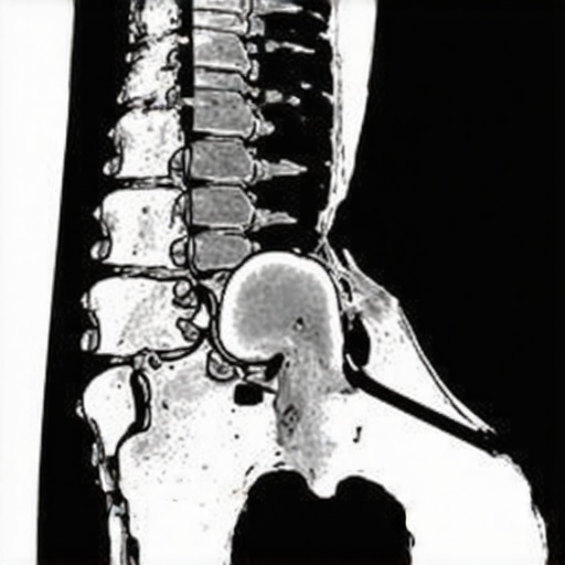
My Journey into Spinal Imaging: A Personal Reflection
As someone who has navigated various orthopedic concerns, I remember the first time I was scheduled for a spinal scan. The uncertainty about what to expect was almost as nerve-wracking as the pain I was experiencing. Over the years, I’ve come to appreciate how important these diagnostic imaging procedures are in pinpointing spinal issues accurately, enabling effective treatment plans.
Understanding the Role of Diagnostic Imaging in Spinal Health
When my doctor recommended a spinal scan, I realized it was a crucial step toward understanding my condition better. These imaging tests, such as MRI or CT scans, provide detailed visuals of the spine, helping to identify herniated discs, nerve compression, or other structural issues. For many patients, including myself, knowing what’s involved demystifies the process and eases anxiety.
What Does a Spinal Scan Entail? My Personal Experience
On the day of my scan, I was instructed to change into a hospital gown and lie still on a narrow table that slides into the MRI machine. The experience was surprisingly comfortable—much better than I anticipated. The procedure took about 30-45 minutes, during which I was asked not to move. The technician explained that the scan produces cross-sectional images that help my orthopedic specialist assess the condition of my spine with exceptional clarity.
How Are the Results Interpreted? A Glimpse into the Diagnostic Process
Once the scan was complete, I waited a few days for my doctor to review the images. The radiologist’s report highlighted areas of concern, which my orthopedic surgeon used to develop a tailored treatment plan. This collaborative approach underscores the significance of high-quality diagnostic imaging in achieving better outcomes, especially for complex spinal conditions.
Why Is Choosing a Skilled Facility in NJ So Important?
In my research, I discovered that the quality of the imaging center can significantly impact the accuracy of the diagnosis. Reputable facilities equipped with advanced MRI machines and experienced radiologists, like those in NJ, ensure precise results. For more insights, you can visit top NJ spine specialists.
If you’re considering a spinal scan, I recommend consulting with an orthopedic expert who understands the nuances of spinal diagnostics. And if you’re curious about other non-invasive treatments, check out minimally invasive back pain treatments.
Have You Had a Spinal Scan? Share Your Experience!
Engaging with others who’ve undergone similar procedures helped me feel more confident. Feel free to comment below or share your story. Remember, understanding what to expect can make all the difference in your journey toward spinal health.
Unveiling the Complexities of Spinal Imaging: Insights from an Orthopedic Specialist
As an orthopedic professional deeply involved in diagnosing and treating spinal conditions, I recognize the importance of precise imaging techniques in crafting effective treatment plans. Beyond standard MRI and CT scans, advanced imaging modalities now offer unparalleled clarity, enabling us to detect subtle anomalies that might otherwise go unnoticed. These innovations are especially crucial in complex cases such as multi-level herniations or atypical nerve compressions, where traditional scans might fall short.
Choosing the Right Imaging Technology for Specific Spinal Concerns
For patients with suspected soft tissue injuries or nerve involvement, high-resolution MRI with contrast enhancement can delineate nerve roots and detect inflammation with exceptional detail. Conversely, CT scans excel in evaluating bony structures, fractures, or post-surgical changes. As a trusted NJ-based specialist, I emphasize the importance of customizing imaging approaches to individual conditions, ensuring the most accurate diagnostics. You can learn more about top NJ spine specialists who utilize these cutting-edge techniques here.

Interpreting Imaging Results: A Critical Step Toward Precision Treatment
The interpretation of spinal images requires a nuanced understanding of anatomy and pathology. For instance, identifying a small disc protrusion pressing on nerve roots demands experience and attention to detail. As specialists, we cross-reference imaging findings with clinical symptoms, ensuring a comprehensive diagnosis. Furthermore, emerging technologies like 3D reconstructions and functional MRI are expanding our ability to visualize complex spinal dynamics, leading to more targeted interventions.
How Imaging Enhances Patient Outcomes and Treatment Planning
Accurate diagnostics inform decisions between conservative management and surgical intervention. For example, detailed imaging can reveal nerve compression severity, guiding whether to pursue minimally invasive procedures or consider fusion surgery. The integration of imaging results with other assessments, such as physical exams and patient history, embodies the holistic approach we advocate for optimal outcomes.
What Are the Latest Advances in Spinal Imaging That Every Expert Should Know?
Recent developments like diffusion tensor imaging (DTI) and upright MRI scans provide functional insights into spinal cord and nerve health, even in weight-bearing positions. These innovations can detect early degenerative changes or nerve entrapments that static images might miss. Staying updated on these advancements is vital for orthopedic specialists committed to delivering cutting-edge care. For a comprehensive understanding of innovative imaging modalities, visit latest advancements in orthopedic imaging.
Empowering Patients Through Knowledge and Collaboration
Educating patients about the significance of advanced imaging fosters trust and compliance. Explaining how these diagnostics guide personalized treatment plans can alleviate anxiety and set realistic expectations. I encourage patients to ask questions and share their experiences, as this collaborative approach enhances overall care. If you’re considering diagnostic imaging, consulting with a reputable NJ orthopedic specialist ensures access to state-of-the-art technology and expert interpretation.
Have You Experienced Cutting-Edge Spinal Imaging? Share Your Insights!
Engaging with others who’ve undergone advanced diagnostics can provide valuable perspectives. Feel free to comment below or share your story. Remember, understanding the nuances of spinal imaging is a cornerstone of effective treatment and recovery. For more insights into comprehensive orthopedic diagnostics and treatments, explore our related articles or reach out through contact us.
My Evolving Understanding of Spinal Imaging: Personal and Professional Reflections
Over the years, my journey with spinal imaging has been both personal and professional. Initially, I approached these diagnostic procedures with a mix of curiosity and caution, often overwhelmed by the complex images and technical jargon. However, as I gained more experience, I realized the profound impact that high-quality imaging has on diagnostic accuracy and subsequent treatment outcomes. This awakening has deepened my appreciation for the advancements in imaging technology, especially in a state like New Jersey where cutting-edge facilities are accessible and integral to patient care.
The Nuances of Choosing the Appropriate Imaging Modality
From my perspective, selecting the right imaging modality is akin to choosing the right tool for a delicate craft. For soft tissue assessment, MRI with contrast can reveal nerve inflammation and subtle disc protrusions that might be missed on a standard scan. Conversely, CT scans excel in evaluating osseous structures, making them indispensable for fracture assessment or post-operative evaluation. Recognizing these nuances has been pivotal in my practice, ensuring that each patient receives tailored diagnostics that inform precise treatment strategies. I often recommend consulting top NJ specialists who leverage these advanced techniques, as their expertise can make a significant difference in outcomes here.
Interpreting the Complex Language of Imaging: A Personal Challenge
One of the most intellectually demanding aspects of my work is deciphering the language of imaging reports. Terms like “disc extrusion” or “nerve root impingement” require not only understanding their anatomical implications but also correlating them with clinical symptoms. This process is akin to translating a foreign language—requiring both technical knowledge and clinical intuition. Recent innovations like 3D reconstructions and functional MRI have been instrumental in bridging this gap, offering dynamic insights into spinal function that static images cannot provide see latest advancements. My goal is always to integrate these technological insights into comprehensive patient care, emphasizing personalized treatment plans.
The Power of Advanced Imaging in Complex Cases
In my practice, I have encountered cases where traditional imaging fell short, especially in diagnosing multi-level herniations or atypical nerve compressions. Here, advanced modalities like diffusion tensor imaging (DTI) have been transformative. DTI allows us to visualize nerve fiber tracts with remarkable detail, enabling earlier detection of degenerative changes and more targeted interventions. This technological leap underscores the importance of continuous education and adaptation in orthopedic care, particularly in a competitive and innovative environment such as NJ. For those seeking expert opinions, exploring the profile of top NJ spine specialists can be enlightening here.
Reflecting on Patient-Centered Care and Technological Advances
Ultimately, my professional philosophy revolves around patient-centered care—using every available tool to craft the most effective, least invasive treatments. Sharing these insights helps demystify the complex world of spinal diagnostics and empowers patients to make informed decisions. I invite those who have experienced advanced spinal imaging to share their stories or ask questions below. Engaging in these discussions not only enriches our collective knowledge but also reinforces the importance of precision medicine in orthopedics.
Exploring the Future: The Boundaries of Spinal Imaging Innovation
The future of spinal imaging is poised for exciting breakthroughs. Emerging technologies like upright MRI scans in weight-bearing positions and advanced functional imaging will further refine our understanding of spinal biomechanics. These tools promise earlier detection of degenerative changes and more personalized treatment pathways. Staying abreast of these developments is essential for any orthopedic professional committed to excellence. For the most current insights, I recommend visiting latest innovations in orthopedic imaging.
Integrating Functional MRI for Dynamic Spinal Assessment
One of the most groundbreaking developments in my professional journey has been the incorporation of functional MRI (fMRI) into diagnostic protocols. Unlike conventional static imaging, fMRI captures real-time spinal cord and nerve activity, providing insights into how degenerative changes influence nerve function during movement or load-bearing scenarios. This modality has been particularly invaluable in diagnosing conditions like early-stage degenerative disc disease or subtle nerve entrapments that traditional MRI might miss. As highlighted by the research from the University of California, these advanced techniques enhance our ability to tailor interventions precisely, ultimately improving patient outcomes through targeted therapies.
Harnessing 3D Reconstruction for Surgical Precision
Beyond 2D cross-sectional images, 3D reconstructions have revolutionized my approach to complex spinal cases. They allow for meticulous preoperative planning, enabling us to visualize the exact spatial relationships of herniations, nerve roots, and bony structures. This clarity reduces intraoperative surprises and enhances surgical accuracy, especially in minimally invasive procedures. Moreover, 3D models facilitate patient education, helping individuals understand their condition better, which fosters trust and compliance. Integrating these models into our practice underscores a commitment to precision medicine, a principle I deeply value in my ongoing efforts to deliver superior care.
What Are the Latest Advances in High-Resolution Nerve Imaging?
High-resolution nerve imaging, particularly diffusion tensor imaging (DTI), has opened new horizons in diagnosing nerve injuries and entrapments. DTI maps nerve fiber tracts with remarkable detail, allowing us to detect early signs of nerve degeneration or subtle compressions that static images might overlook. This technology is especially beneficial in cases where symptoms are disproportionate to imaging findings, guiding us toward more effective interventions. Staying abreast of such innovations is crucial for orthopedic specialists dedicated to comprehensive care. For an in-depth look at how these advancements are shaping our field, you can explore authoritative sources like the Journal of Orthopedic Research, which regularly publishes pioneering studies.
Engage with Cutting-Edge Knowledge to Elevate Your Treatment Strategies
If you’re a fellow clinician or a patient eager to understand these sophisticated diagnostic tools, I invite you to connect and share insights. Embracing technological innovation is vital in our pursuit of excellence. Feel free to reach out through our contact page for personalized discussions or to schedule a consultation. Your journey toward optimal spinal health deserves the most advanced, evidence-based approaches—let’s explore them together.
Things I Wish I Knew Earlier (or You Might Find Surprising)
1. The Power of Precise Imaging
Looking back, I realize how much clearer my diagnosis became once I understood the importance of high-quality spinal imaging. Early on, I underestimated how detailed images like MRI and CT scans could reveal subtle issues that might be missed on standard X-rays. This knowledge transformed my approach to treatment and recovery.
2. The Nuance of Choosing the Right Modality
Choosing between MRI, CT, or advanced techniques like DTI isn’t a one-size-fits-all decision. Each has its strengths—MRI for soft tissues, CT for bones—and knowing when to use each can make a big difference in outcomes. I wish I had grasped these nuances sooner, as it would have saved time and anxiety.
3. The Role of Advanced Technology
Emerging tools like functional MRI and 3D reconstructions are game changers. They provide dynamic insights, especially in complex cases, helping specialists craft more targeted treatments. Discovering these innovations made me appreciate how technology continuously elevates patient care.
4. The Value of Collaboration
Interpreting imaging results isn’t just about reading scans; it’s about collaboration between radiologists, orthopedic surgeons, and patients. When everyone speaks the same language and understands the images, treatment plans are more precise and effective. I’ve seen firsthand how teamwork enhances recovery.
5. Trust in Reputable Facilities
The quality of imaging centers can vary widely. I’ve learned to seek out NJ-based facilities with advanced equipment and experienced radiologists for the most accurate results. Trustworthy centers can truly impact your journey towards spinal health.
6. The Future Is Exciting
The future of spinal imaging holds promise with innovations like upright MRI and real-time nerve imaging. Staying informed about these developments ensures I can offer my patients the best care possible. It’s an exciting time to be in this field.
Resources I’ve Come to Trust Over Time
1. The Journal of Orthopedic Research
This peer-reviewed journal provides cutting-edge studies on imaging technology. It’s been invaluable for understanding the latest innovations and clinical applications, making it a must-read for professionals and curious patients alike.
2. The American Journal of Roentgenology
Specializing in radiology, this resource offers detailed insights into imaging techniques and advancements. It’s helped me stay updated on best practices and emerging modalities.
3. The National Institute of Biomedical Imaging and Bioengineering (NIBIB)
As a trusted authority, NIBIB shares accessible research and breakthroughs in medical imaging. I recommend exploring their resources to anyone interested in the future of diagnostic tools.
4. Local NJ Imaging Centers’ Publications
Many reputable NJ facilities publish educational content and case studies. These resources are practical and relevant, especially for patients seeking nearby expert care.
Parting Thoughts from My Perspective
Reflecting on my journey through spinal imaging, I realize how vital these diagnostic tools are in shaping effective treatment and recovery. The combination of technological innovation, expert interpretation, and patient-centered care creates a powerful synergy. If you’re navigating spinal issues or simply curious about the process, I encourage you to seek out reputable NJ specialists and stay informed about advancements. Sharing experiences and questions can empower us all on this journey toward better spinal health. If this resonated with you, I’d love to hear your thoughts or stories—feel free to comment below or reach out through our contact page. Remember, knowledge is the first step toward healing, and you don’t have to face it alone.

Reading this post truly resonated with my own experience undergoing spinal imaging in NJ. I was initially quite anxious about the process, especially about what the results might reveal. However, the detailed explanation about the procedure and importance of selecting a skilled facility helped ease my fears. I particularly appreciated the emphasis on different imaging modalities like MRI and CT scans — knowing when each is most effective can be quite confusing for patients. In my case, an advanced MRI with contrast provided the clarity needed to diagnose a nerve impingement I had been battling for months. It’s impressive how technology like 3D reconstructions and functional MRI now enhances our understanding and treatment planning. Has anyone here had success with newer imaging techniques like DTI or upright MRI scans? I believe staying informed about these innovations can really make a difference in managing complex cases effectively.