Getting Ready for My Orthopedic MRI: A Personal Perspective
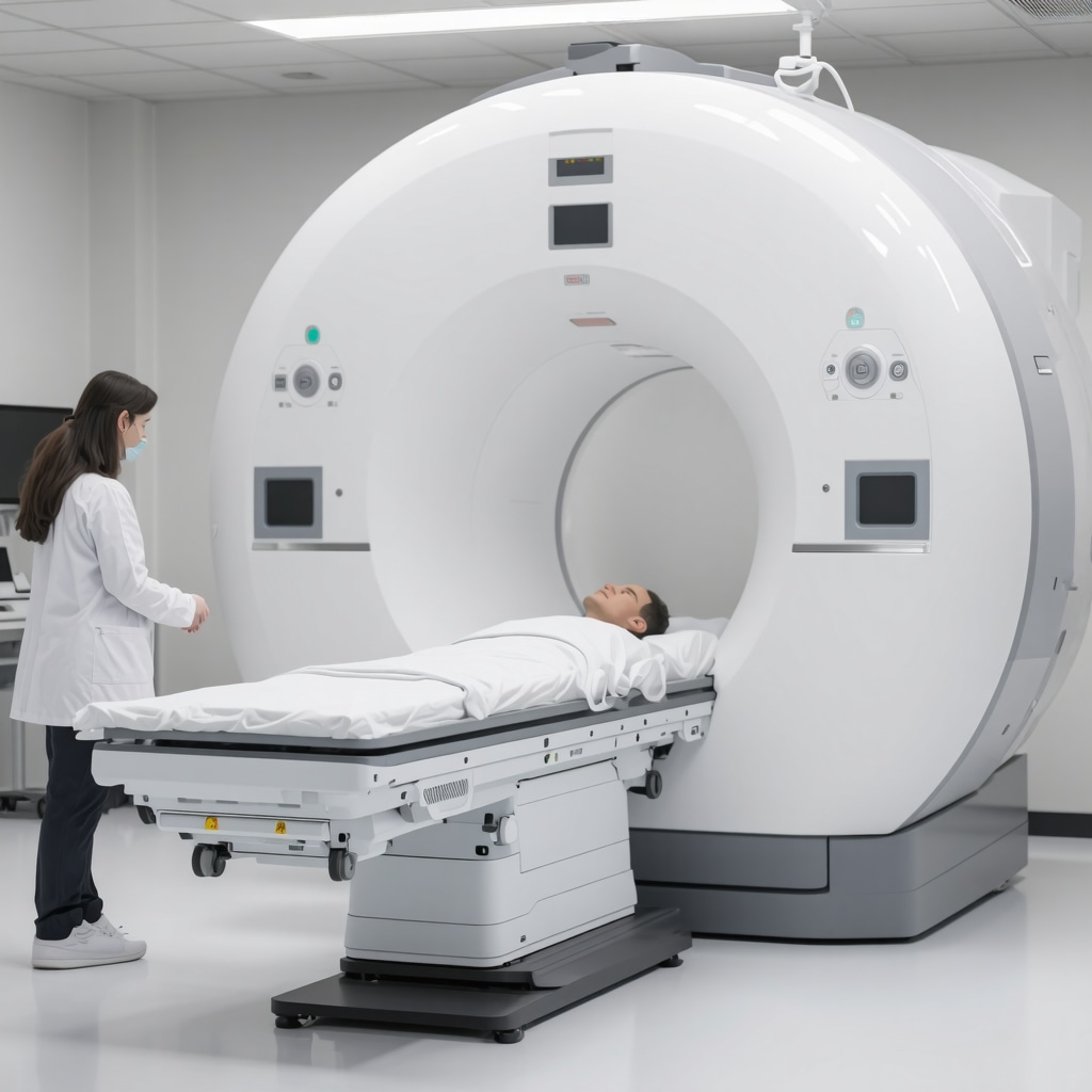
When I first faced the prospect of needing an orthopedic MRI in 2025, I was filled with a mix of curiosity and apprehension. Having experienced chronic back pain for years, I knew that accurate imaging was crucial for proper diagnosis and effective treatment. My journey into preparing for this diagnostic test was not just about showing up at the imaging center; it was about understanding what to expect and how to make the process as smooth as possible.
Understanding the Role of Imaging in Orthopedics
In my experience, MRI and other imaging techniques like X-rays play a vital role in diagnosing complex musculoskeletal issues. As shared by leading orthopedic radiologists, MRI provides detailed images of soft tissues, which are essential when assessing ligament, cartilage, or disc problems. I learned that knowing the differences between MRI and X-ray helps in understanding how your doctor will evaluate your condition. For instance, MRI is often preferred for soft tissue injuries, while X-ray is more common for bone issues.
How I Prepared for My MRI Appointment
Preparation was key. I was advised to wear comfortable, loose clothing without metal fasteners and to avoid wearing jewelry. I also made sure to inform the staff if I had any implants or metallic devices. A helpful tip was to stay well-hydrated and avoid caffeine before the scan, which can sometimes interfere with imaging quality. Most importantly, I read that bringing a list of current medications and medical history can streamline the process and ensure accurate results. For more detailed tips, I referred to authoritative sources like the American College of Radiology’s guidelines.
What Does the MRI Experience Feel Like?
The actual procedure was surprisingly comfortable. The machine is quite loud, so I was given headphones to block out the noise. The technician explained that the scan would last about 30-45 minutes and that I should remain still to get clear images. I appreciated the reassurance and clear communication from the staff, which made the experience less intimidating. Remember, if you have anxiety or claustrophobia, discussing options with your doctor beforehand can be very helpful.
Why Accurate Preparation Matters
Accurate preparation not only improves the quality of the images but also speeds up the diagnosis process. This means quicker treatment planning and better outcomes. If you’re like me and value clarity and precision, taking the time to understand what’s involved can make a significant difference. Also, don’t forget to follow up with your doctor after the imaging to discuss the results and next steps.
How do I decide between MRI and other imaging techniques for my orthopedic issues?
This is a common question I had. Generally, your doctor will recommend the most suitable imaging based on your symptoms and medical history. For soft tissue injuries, MRI is often the best choice, while X-rays are more suitable for detecting fractures or bone alignment issues. Consulting with an experienced orthopedic spine specialist can help you navigate these choices. For example, you might explore options at trusted clinics like top orthopedic spine specialists.
If you’re preparing for an upcoming MRI, I encourage you to share your questions or experiences in the comments below. Sharing tips and insights can help others feel more confident and prepared for their diagnostics.
Deciphering Orthopedic Imaging: Choosing the Best Technique for Your Spine Issues
As an experienced orthopedic specialist, I often encounter patients overwhelmed by the variety of imaging options available for diagnosing spinal conditions. Understanding the nuances between MRI, X-ray, CT scans, and other modalities is crucial for tailored treatment plans. Each imaging technique offers unique insights, and selecting the right one depends on the specific clinical scenario, patient history, and suspected pathology.
Why MRI Remains the Gold Standard for Soft Tissue Evaluation
Magnetic Resonance Imaging (MRI) is unparalleled in its ability to visualize soft tissues such as discs, ligaments, nerve roots, and muscles. This makes MRI the preferred choice when evaluating herniated discs, spinal stenosis, or nerve impingements. For instance, if a patient presents with radiculopathy, an MRI can reveal nerve compression with remarkable clarity, guiding precise interventions.
When to Opt for X-ray or CT Scans in Spine Assessment
X-rays are excellent for assessing bony alignment, fractures, or degenerative changes like osteoarthritis. They are quick, accessible, and involve less cost, making them ideal for initial assessments or follow-up. Conversely, Computed Tomography (CT) scans provide detailed bone imaging and are useful when evaluating complex fractures or planning surgical interventions. For example, a suspected vertebral fracture often warrants a CT scan for comprehensive visualization.
How Advanced Imaging Enhances Treatment Planning
Incorporating advanced imaging techniques allows orthopedic surgeons to craft more effective treatment strategies. For example, diffusion-weighted MRI can detect subtle nerve injuries, while 3D reconstructions from CT scans can assist in surgical planning. Consulting trusted sources like the American College of Radiology underscores the importance of choosing appropriate imaging modalities to improve diagnostic accuracy and patient outcomes. For further insights, visit orthopedic radiology centers.
Balancing Risks and Benefits of Imaging Procedures
While MRI and other imaging techniques are invaluable, they are not without considerations. MRI is contraindicated in patients with certain metallic implants or devices. X-rays and CT scans involve exposure to radiation, so their use should be judicious, especially in younger populations. As an expert, I emphasize the importance of collaborative decision-making with radiologists and specialists to optimize safety and diagnostic yield.
Can you tell me about your experience with different imaging modalities and how they influenced your treatment decisions?
Sharing real-world experiences helps demystify the process and empowers patients and practitioners alike. If you’re preparing for an orthopedic evaluation, consider discussing your imaging options with your doctor to understand the rationale behind each choice. For personalized guidance, exploring reputable clinics and specialists, such as top orthopedic spine specialists, can make a significant difference.
For more tips on navigating your orthopedic care journey, feel free to leave a comment or share this article with others seeking clarity on spinal imaging techniques.
Reflecting on the Nuances of MRI Preparation and Personal Experiences
As I continue to navigate the complex world of orthopedic imaging, I realize that each patient’s journey is unique and layered with subtle considerations that can significantly impact diagnostic accuracy. My own experiences have shown me that preparation goes beyond simple guidelines—it’s about understanding how personal health history, emotional state, and even environmental factors influence the imaging process. For instance, I learned that mental preparedness and relaxation techniques can reduce anxiety during the scan, leading to better cooperation and clearer images. Engaging with patients on these nuanced levels fosters a more holistic approach to care.
Choosing the Optimal Imaging Technique: Beyond the Basics
In my practice, I’ve seen how the decision between MRI, X-ray, or CT scans hinges on more than just clinical indications. It involves considering the patient’s lifestyle, previous medical procedures, and even their future treatment plans. For example, a patient with a metallic implant might benefit from a low-dose CT scan to avoid MRI contraindications, whereas soft tissue evaluation might necessitate an MRI despite the presence of some implants, provided they are MRI-compatible. This layered decision-making process underscores the importance of personalized care, which is supported by authoritative sources like the American College of Radiology, emphasizing tailored imaging strategies.
The Psychological Aspect: Managing Anxiety and Claustrophobia
One aspect I’ve come to appreciate deeply is the psychological comfort of patients. Even with thorough preparation, some individuals experience claustrophobia or heightened anxiety, which can compromise image quality. I found that discussing these fears openly and exploring options like sedation or open MRI machines can make a difference. Personally, I’ve seen patients benefit from calming techniques or guided imagery, which not only improve their experience but also enhance the reliability of the results. Recognizing and addressing these emotional factors is an integral part of comprehensive orthopedic care.
Expert Insights: Advancing Imaging Technologies and Future Directions
Staying abreast of technological advancements, such as 3D imaging and functional MRI, allows me to envision how future diagnostics might evolve. These innovations promise even more precise visualization of soft tissues and nerve function, revolutionizing treatment planning. I often refer to authoritative sources, like the American College of Radiology, to validate these emerging techniques. For example, diffusion tensor imaging (DTI) is gaining traction for nerve injury assessment, offering insights that were previously unattainable. Embracing these advancements requires continuous learning and adaptability, which I find intellectually stimulating and vital for delivering cutting-edge care.
Inviting Readers to Share and Explore Further
Understanding the intricate layers of orthopedic imaging is a journey that benefits from shared experiences and ongoing dialogue. I encourage readers to reflect on their own experiences with diagnostic imaging and consider how personalized preparation and awareness can improve outcomes. If you’re navigating similar challenges or curious about specific techniques, feel free to share your insights or questions in the comments. Exploring trusted resources like choosing the right orthopedic surgeon can also guide you toward making informed decisions tailored to your needs. Together, we can deepen our understanding and enhance the quality of orthopedic care for all.
Exploring the Nuances of MRI Technology and Its Impact on Treatment Precision
As my experience deepened, I recognized that the evolution of MRI technology has significantly enhanced diagnostic accuracy, especially with the advent of functional MRI and diffusion tensor imaging (DTI). These sophisticated modalities allow us to visualize nerve fiber pathways and detect subtle soft tissue injuries that traditional MRI might miss. For example, DTI has become instrumental in evaluating nerve root impingements, guiding minimally invasive interventions and personalized treatment plans. According to a comprehensive review in the American Journal of Roentgenology, such advancements are revolutionizing how orthopedic specialists approach complex cases, emphasizing the importance of staying updated with technological progress source.
How do these emerging imaging techniques influence surgical decision-making and patient outcomes?
Implementing advanced imaging insights can lead to more targeted interventions, reducing unnecessary procedures and optimizing recovery trajectories. For instance, precise nerve imaging can determine whether conservative management is feasible or if surgical decompression is warranted. I encourage fellow practitioners and patients to engage in discussions about these innovations, as they hold the potential to transform standard care protocols. For those interested in exploring cutting-edge diagnostics further, consulting with top spine specialists, such as leading experts in 2025, can provide valuable insights into personalized treatment pathways.
The Critical Role of Personalized Imaging Strategies in Chronic Spinal Conditions
Every patient’s anatomy and pathology are unique, necessitating tailored imaging approaches. My practice has shifted toward integrating multiple modalities—combining MRI, CT, and occasionally dynamic imaging—to capture a comprehensive picture of spinal biomechanics. For example, in cases of suspected instability, flexion-extension X-rays supplemented by MRI can reveal hidden subluxations and soft tissue compromises. This layered imaging approach allows for more precise surgical planning, especially in complex cases involving degenerative disc disease or spondylolisthesis. It underscores the importance of a multidisciplinary perspective, supported by evidence from institutions like the National Institute of Health, which emphasizes personalized diagnostics for improved outcomes NIH research.
What are the benefits and limitations of combining multiple imaging techniques in complex spinal diagnoses?
While this strategy enhances diagnostic accuracy, it also demands careful interpretation to avoid conflicting findings. It requires collaboration among radiologists, orthopedic surgeons, and physical therapists to synthesize data effectively. I invite colleagues and patients to consider this comprehensive approach, as it often results in more effective, less invasive treatment plans. For those seeking expert guidance, reaching out to multidisciplinary clinics specializing in spinal care can provide access to integrated diagnostic and therapeutic solutions.
Addressing Psychological Barriers to Imaging and Treatment Acceptance
Throughout my journey, I observed that emotional factors such as claustrophobia and anxiety significantly influence patient experiences and diagnostic quality. Incorporating psychological support, relaxation techniques, and open communication has proven beneficial. For example, some patients benefit from guided imagery or sedation, facilitating a calmer, more cooperative environment during scans. Recognizing these barriers is vital, as unresolved anxiety can lead to suboptimal imaging and delayed treatment. The integration of mental health strategies into orthopedic care reflects a holistic approach endorsed by the American Psychological Association, emphasizing the mind-body connection in healing APA studies.
How can practitioners better prepare patients psychologically for advanced imaging procedures?
Proactive communication, empathetic engagement, and offering calming resources can make a significant difference. I recommend providing detailed explanations, virtual tours of the imaging environment, and discussing sedation options when appropriate. Patients who feel informed and supported tend to have better experiences, which ultimately enhances diagnostic accuracy and treatment success. Sharing success stories and practical tips on platforms like our contact page can foster community trust and empowerment.
Things I Wish I Knew Earlier (or You Might Find Surprising)
Preparation Is More Than Just Wearing Loose Clothing
Initially, I thought just avoiding jewelry was enough, but I soon realized that wearing comfortable, loose clothing without any metal fasteners makes a huge difference. It’s amazing how small details can impact the quality of your images and overall experience.
Hydration Really Matters
I didn’t expect that staying well-hydrated before the scan could influence the imaging results. Drinking plenty of water helps your body and can make the process smoother, especially if contrast agents are involved.
Knowing Your Implants Is Crucial
Informing the staff about any implants or metallic devices early on can prevent delays or complications. I learned to keep a list of my medical devices handy—something I wish I did sooner.
Managing Anxiety Can Improve the Experience
My first MRI was a bit intimidating, but practicing relaxation techniques and discussing fears with the technician made a big difference. If you have claustrophobia, exploring options like open MRI or sedation can be worthwhile.
Follow-Up Is Key
After the scan, I realized that understanding the results and discussing next steps with my doctor helps me feel more in control of my treatment journey. Don’t hesitate to ask questions or seek clarifications.
Resources I’ve Come to Trust Over Time
- American College of Radiology: Their guidelines on MRI preparation are comprehensive and reliable. I’ve found their resources helpful for understanding what to expect.
- National Institutes of Health (NIH): Their research articles on imaging techniques deepen my understanding of the latest advancements and how they influence treatment.
- RadiologyInfo.org: A user-friendly site that explains imaging procedures in simple terms. I recommend it to anyone feeling overwhelmed by medical jargon.
- Leading Orthopedic Specialists in 2025: Checking out top clinics through trusted directories has connected me with experienced professionals who prioritize patient education and comfort.
Parting Thoughts from My Perspective
Preparing for an orthopedic MRI in 2025 taught me that small details and honest communication can significantly improve your experience. I’ve learned to be proactive—asking questions, understanding my own health history, and managing my anxiety. These steps not only lead to better images but also foster a sense of confidence and control over my health journey. If you’re about to undergo an MRI, remember that knowledge and preparation are your best allies. If this resonated with you, I’d love to hear your thoughts or tips. Sharing stories helps us all navigate the complexities of orthopedic care with more ease and understanding. Feel free to connect and ask questions—your journey matters, and you’re not alone in it.


Reading this detailed journey into preparing for an orthopedic MRI really resonated with me. I recently went through a similar experience for a knee injury, and I found that staying relaxed and asking questions beforehand made the process much smoother. I also appreciated the emphasis on hydration—something I overlooked and wish I had known earlier. One thing I’ve learned is that bringing a calming playlist or practicing breathing techniques can help ease anxiety during the scan, especially for those who are claustrophobic. Has anyone else found particular strategies effective for managing nervousness during MRIs? Additionally, I wonder how newer imaging technologies like functional MRI or DTI will influence future treatment plans. It’s exciting to think about the possibilities, but I’d love to hear what experiences others have had with these advanced techniques and how they affected their recovery or surgical decisions.