My Journey into the World of Orthopedic MRI & Imaging
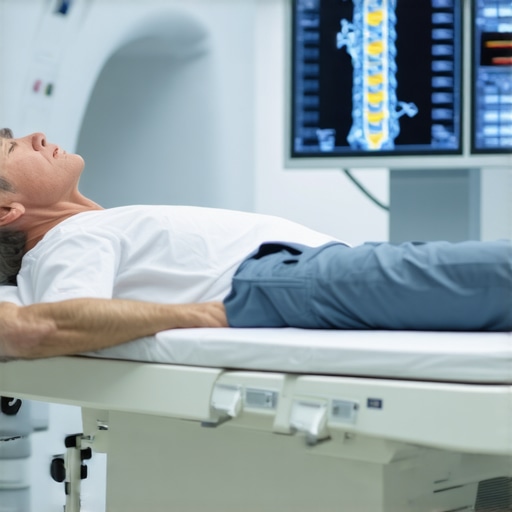
It all started when I experienced persistent back pain that just wouldn’t go away. After a frustrating week of discomfort, I finally decided to consult an orthopedic specialist. During my visit, I was amazed to learn about the vital role that orthopedic MRI & imaging play in diagnosing complex spinal conditions with remarkable accuracy.
Why I Realized Advanced Imaging is a Game Changer
During my consultation, my doctor explained how MRI technology can reveal detailed images of soft tissues, discs, nerves, and bones — far beyond what X-rays can show. This comprehensive view helps in pinpointing the exact source of pain, whether it’s a herniated disc, nerve compression, or other spinal issues. I was surprised to discover that MRI scans are non-invasive and painless, making them a preferred choice for precise diagnostics.
The Personal Impact of Accurate Spinal Diagnostics
After my MRI, I received a clear diagnosis that enabled my doctor to tailor a treatment plan specifically for my condition. This experience underscored the importance of high-quality imaging in avoiding unnecessary surgeries and choosing effective, minimally invasive therapies. For anyone suffering from chronic back or neck pain, understanding how advanced imaging works can be a crucial step toward relief.
How Can MRI & Imaging Improve Your Spinal Health?
From my experience, the benefits of orthopedic MRI & imaging extend beyond diagnosis. They guide treatment options such as targeted physical therapy, epidural injections, or even surgical interventions if necessary. As detailed by the American College of Radiology, MRI provides invaluable insights that help doctors develop comprehensive care strategies (source).
Ever Wondered How MRI Scans Detect Nerve Compression or Disc Herniation?
Understanding the mechanics behind MRI technology can be fascinating. These scans produce cross-sectional images that reveal nerve impingements, disc bulges, and vertebral abnormalities with incredible clarity. This allows surgeons and physiotherapists to plan precise interventions, leading to better outcomes and quicker recovery times.
If you’re considering an MRI for your spinal concerns, I highly recommend discussing with your healthcare provider about the latest imaging options available. It might just be the key to unlocking your path to recovery. Feel free to share your experiences or ask questions in the comments below — I’d love to hear your story!
The Critical Role of MRI & Imaging in Modern Orthopedics
Orthopedic MRI & imaging techniques have revolutionized how specialists diagnose and treat spinal conditions. These advanced tools provide detailed insights that are often unattainable through traditional X-rays, capturing soft tissues, nerves, and discs with remarkable clarity. As a result, clinicians can pinpoint issues like nerve compression, disc herniation, or ligament tears with high accuracy, leading to more targeted and effective treatments.
Choosing the Right Imaging Modality for Your Spinal Condition
While MRI is the gold standard for soft tissue evaluation, other imaging options like CT scans or myelograms can be valuable depending on specific circumstances. For instance, CT scans offer excellent bone detail, making them suitable for assessing complex fractures or bony abnormalities. However, MRI’s ability to visualize soft tissues makes it indispensable for diagnosing herniated discs or nerve impingements. Consulting with your orthopedic specialist can help determine the most appropriate imaging approach for your needs.
Advances in MRI Technology: What’s New in 2025?
Recent innovations, such as high-field MRI machines and functional imaging, have enhanced diagnostic precision. These developments allow for detailed visualization of nerve function and blood flow, providing deeper insights into spinal health. Moreover, faster scan times reduce patient discomfort and improve clinic throughput. Staying informed about these technological advancements ensures that patients receive the most accurate diagnostics possible.
How Can Imaging Inform Your Treatment Plan?
High-quality imaging results are integral in crafting personalized treatment strategies. Whether it’s guiding minimally invasive procedures like epidural injections or planning complex surgeries, detailed scans ensure interventions target the root cause of pain. For example, understanding the exact location and extent of nerve compression can determine whether a patient benefits from conservative therapy or needs surgical intervention.
The Expert’s Perspective: Why Reliable Imaging Matters
As a seasoned orthopedic specialist, I emphasize that accurate imaging not only facilitates precise diagnosis but also enhances patient safety. It minimizes unnecessary procedures and helps set realistic recovery expectations. According to the American College of Radiology, comprehensive imaging is vital in developing effective, evidence-based care pathways (source).
Are You Aware of How MRI & Imaging Can Detect Early Signs of Spinal Degeneration?
Early detection of degenerative changes, such as disc dehydration or ligament thinning, can significantly influence treatment outcomes. Advanced MRI techniques enable clinicians to identify these subtle alterations before they cause severe pain or disability, allowing for proactive management. This proactive approach not only preserves spinal function but can also delay or prevent the need for invasive procedures.
If you’re considering imaging options for your spinal health, I encourage you to discuss the latest innovations with your healthcare provider. Proper diagnostics are the cornerstone of effective treatment, and understanding your options empowers you to make informed decisions. Feel free to share your experiences or ask questions in the comments — your insights can help others navigate their spinal health journey!
Delving Deeper: The Nuances of MRI Interpretation in My Practice
As I have gained more experience in orthopedic imaging, I’ve come to appreciate that MRI isn’t just about capturing clear images; it’s about interpreting subtle nuances that can dramatically influence treatment paths. The differentiation between a minor disc bulge and a significant herniation, for example, often hinges on minute details that require a keen eye and profound understanding of spinal anatomy. This meticulous analysis reminds me of the importance of continuous learning and staying abreast of technological advancements, such as functional MRI techniques, which provide insights into nerve activity and blood flow, elevating diagnostic precision even further (source).
The Personal Journey: From Imaging to Empathy
My journey into the world of MRI has reinforced the vital connection between technological expertise and compassionate patient care. When I review scans, I don’t see just images; I see stories of pain, resilience, and hope. Understanding the complex interplay of soft tissues, nerves, and bones allows me to communicate more effectively with patients, helping them grasp their conditions beyond the technical jargon. This empathy-driven approach underscores that advanced imaging is not merely a diagnostic tool but a bridge to trust and reassurance.
What Are the Limitations and Ethical Considerations of MRI?
While MRI technology has transformed orthopedic diagnostics, it’s essential to recognize its limitations. For instance, incidental findings—such as benign cysts or anatomical variants—can sometimes lead to unnecessary anxiety or interventions. Balancing the benefits of detailed imaging with ethical considerations about overdiagnosis and patient well-being is a nuanced aspect of my practice. Staying informed through authoritative guidelines, such as those from the American College of Radiology, helps ensure I provide responsible and patient-centered care (source).
How Can Orthopedic Professionals Continue to Evolve with Rapid Imaging Advances?
Continuous education is paramount. Attending specialized workshops, engaging with peer-reviewed research, and exploring emerging technologies like AI-enhanced image analysis are ways I stay ahead. AI, in particular, holds promise for detecting subtle abnormalities that might elude even experienced eyes, potentially reducing diagnostic errors and improving outcomes (source). Embracing these innovations while maintaining a patient-focused philosophy is the key to advancing in this field.
If you’re passionate about spinal health or considering imaging options, I encourage you to share your experiences or pose questions. Understanding the deeper layers of MRI’s capabilities can empower you to make more informed decisions. Feel free to explore related topics like post-surgical rehabilitation or how to select the right specialist. Your journey toward better spinal health is uniquely personal, and I’m here to support that path with insights grounded in both technology and compassion.
The Evolution of MRI Technology: Pushing the Boundaries of Spinal Diagnostics
As I continue to deepen my understanding of orthopedic imaging, I’ve witnessed firsthand how the rapid advancements in MRI technology are transforming patient care. The integration of high-field magnets, such as 7 Tesla MRI machines, has significantly improved resolution, allowing for visualization of minute anatomical structures that were previously elusive. This leap in imaging clarity enables clinicians to detect early degenerative changes—like subtle ligament tears or microdisplacements—before they progress into more debilitating conditions. Such early detection is crucial in formulating proactive treatment plans that preserve spinal integrity and function (source).
How Are Functional MRI and Diffusion Tensor Imaging Shaping Spinal Care?
Functional MRI (fMRI) and Diffusion Tensor Imaging (DTI) are emerging modalities that offer insights beyond static anatomy. These techniques assess nerve activity, blood flow, and neural pathways, providing a dynamic perspective on spinal cord and nerve health. For example, DTI can map nerve fiber tracts, helping to identify microstructural impairments that correlate with clinical symptoms like radiculopathy or myelopathy. Incorporating these advanced imaging techniques into my practice has enhanced my ability to develop targeted, minimally invasive interventions, reducing recovery times and improving outcomes.
The Nuances of MRI Interpretation: From Image to Insight
Interpreting MRI scans transcends technical skill; it demands a nuanced understanding of spinal biomechanics and pathology. Differentiating between a benign annular tear and a symptomatic disc herniation often hinges on subtle signal changes and anatomical orientation. My experience underscores that continuous education—attending specialized workshops and engaging with the latest research—is vital in mastering these nuances. For instance, recognizing the significance of Modic changes in vertebral endplates can influence whether conservative management or surgical intervention is appropriate (source).
What Ethical Challenges Do We Face with Increasing MRI Capabilities?
With greater imaging detail comes the risk of incidental findings—benign anomalies that may lead to unnecessary anxiety or interventions. Navigating these ethical considerations requires a balanced approach, emphasizing patient education and shared decision-making. It’s essential to communicate that not all abnormalities warrant treatment, aligning with guidelines from authoritative bodies like the American College of Radiology. My commitment is to ensure that technological capabilities serve the patient’s best interests without overmedicalization.
Embracing AI and Machine Learning in Orthopedic Imaging
The integration of Artificial Intelligence (AI) and machine learning algorithms is poised to revolutionize MRI diagnostics. These tools can assist in detecting subtle abnormalities, quantifying degeneration, and predicting disease progression with unprecedented accuracy. From my perspective, leveraging AI not only enhances diagnostic precision but also streamlines workflow, allowing for more personalized patient care. As research progresses, I anticipate AI will become an indispensable component of orthopedic imaging, guiding treatment decisions with data-driven confidence (source).
Deepening Patient-Doctor Collaboration Through Advanced Imaging
Ultimately, the goal of integrating cutting-edge MRI technology is to foster a deeper partnership with patients. Sharing detailed, comprehensible images helps demystify complex spinal conditions, empowering patients to participate actively in their care. My approach involves translating sophisticated imaging findings into relatable language, emphasizing that technology is a tool to enhance understanding and shared decision-making. This collaborative process is essential for achieving optimal results and sustained spinal health.
If you’re eager to explore how these technological innovations can improve your spinal health or have questions about the latest imaging options, I invite you to reach out through my contact page. Embracing these advancements is not just about better diagnostics—it’s about transforming patient care and outcomes for a healthier future.
Things I Wish I Knew Earlier (or You Might Find Surprising)
Hidden Depths of Soft Tissues
When I first learned about MRI technology, I was amazed that it could reveal soft tissues with such clarity. It’s like having a window into the body’s hidden corners, uncovering issues that X-rays simply can’t show. Discovering this early on changed my perspective on diagnostics and emphasized the importance of advanced imaging in accurate diagnosis.
The Power of Early Detection
I realized that MRI scans can detect early signs of degeneration, such as subtle disc dehydration or ligament thinning, before symptoms become severe. For patients, this means proactive treatment options that can preserve spinal health longer, which I find incredibly empowering and hopeful.
Beyond Just Diagnosing
Advanced imaging isn’t only about identifying problems; it’s also about guiding personalized treatment plans. Whether it’s deciding between physical therapy, injections, or surgery, high-quality MRI results serve as a roadmap that can significantly influence outcomes.
The Limitations and Ethical Considerations
While MRI technology is remarkable, I’ve learned that incidental findings can sometimes lead to unnecessary worries or procedures. Balancing technological capabilities with ethical responsibility is crucial, reminding us that not every anomaly warrants intervention.
The Future Is Bright with AI
The integration of AI and machine learning into MRI analysis is an exciting frontier. These tools can detect minute abnormalities more precisely and help prevent diagnostic errors, making spinal care even more effective in the coming years.
Resources I’ve Come to Trust Over Time
- American College of Radiology (ACR): Their guidelines and research have deepened my understanding of imaging best practices. It’s a go-to resource for evidence-based standards.
- PubMed: The vast database of medical research articles helps me stay updated on the latest advancements in MRI technology and spinal diagnostics.
- Journal of Magnetic Resonance Imaging (JMRI): Specialized articles here provide detailed insights into cutting-edge MRI techniques that I find invaluable.
Parting Thoughts from My Perspective
Exploring the world of orthopedic MRI & imaging has truly transformed how I approach spinal health. I believe that understanding the nuances of advanced diagnostics can empower patients and clinicians alike, leading to better outcomes and more personalized care. If this article resonated with you, I’d love to hear your thoughts. Feel free to share your experiences or questions in the comments — your story could inspire someone else on their journey toward spinal wellness.