Ever Wondered What Really Goes Into a Work Injury Orthopedic Evaluation?
Picture this: You’ve just had a mishap on the job, and suddenly, you’re navigating a maze of medical jargon and appointments. It’s overwhelming, right? Well, don’t worry—there’s a method to this madness, and understanding the essential steps in an orthopedic evaluation can be your ticket to faster recovery and better care.
The First Step: The Art of Listening—More Than Just a Conversation
Think of your orthopedic doctor as a detective. They start by gathering clues—your story. This involves a detailed history of your injury, previous illnesses, and how the pain impacts your daily life. Did you know that a well-conducted history can reveal underlying issues that might complicate recovery? For example, if you’ve had prior spinal problems, your treatment plan might need a different approach. As the top spine specialists emphasize, a thorough evaluation hinges on comprehensive information.
Physical Examination: The Body’s Detective Work
Next, it’s time for the physical exam—your doctor’s chance to perform a meticulous assessment. This includes checking your range of motion, muscle strength, and neurological function. Ever wondered why they tap, press, and move your limbs? It’s to pinpoint the exact location and nature of your injury. This step is crucial because it guides the diagnostic process, ensuring no detail is overlooked.
Imaging and Tests: Peering Inside Without Opening You Up
Sometimes, words and physical exams aren’t enough. That’s where imaging tests like X-rays, MRIs, or CT scans come into play. These tools give doctors a detailed look inside your body, confirming or ruling out suspected injuries. For instance, an MRI can reveal herniated discs that might be pressing on nerves, a common culprit in work-related back pain. If you’re curious about non-invasive options, check out non-surgical care strategies.
Why It Matters: The Power of an Accurate Evaluation
Getting this evaluation right isn’t just about diagnosing; it’s about setting the stage for effective treatment. An accurate assessment helps avoid unnecessary procedures, speeds up recovery, and ensures you’re on the right path. As one expert notes, “A detailed work injury orthopedic evaluation is the cornerstone of personalized care” (source).
Are You Ready to Take Control of Your Recovery?
If you’ve experienced a work injury, understanding these steps can empower you to advocate for yourself. Remember, a comprehensive evaluation is your first step toward effective care. Share your thoughts or experiences in the comments below—your story might help someone else navigate their recovery journey.
How Can a Deep Understanding of Your Orthopedic Evaluation Accelerate Your Recovery?
Imagine this: after an on-the-job mishap, you’re faced with a barrage of medical appointments and complex terminology. But what if you could demystify the process? A comprehensive orthopedic evaluation isn’t just a bureaucratic hurdle—it’s your gateway to personalized and effective treatment. By understanding the nuanced steps involved, you empower yourself to collaborate better with healthcare providers and speed up your return to health.
Beyond Surface-Level Assessments: The Nuanced Art of Medical History
One might wonder, why does your doctor spend so much time delving into your past injuries, daily routines, and even your pain patterns? The answer lies in the detailed history taking, which acts as a roadmap for diagnostic accuracy. A well-conducted history can reveal underlying issues that influence treatment, such as previous spinal conditions or repetitive strain injuries. As the top orthopedic spine specialists highlight, this step is crucial in tailoring interventions that are truly effective.
The Significance of a Thorough Physical Exam
Next comes the physical examination—your doctor’s chance to perform a meticulous, hands-on assessment. They evaluate your range of motion, muscle strength, reflexes, and neurological responses. Ever wondered why they press and tap on different parts of your body? These maneuvers help localize the injury and determine its severity. This process is vital; an incomplete exam can lead to misdiagnosis or overlooked issues, delaying recovery. For example, identifying nerve impingement early can be the difference between a quick recovery and months of pain.
Deciphering Internal Clues: The Role of Imaging and Diagnostic Tests
When the physical exam raises questions, imaging studies like X-rays, MRIs, or CT scans step in to provide a detailed view inside your body. These tools are invaluable in confirming suspected injuries or ruling out serious conditions. For instance, an MRI can reveal herniated discs pressing on nerves—common culprits behind work-related back pain. If you’re curious about less invasive options, explore non-surgical care strategies that might be suitable for your situation.
The Power of Accurate Evaluation in Personalized Treatment
An accurate and comprehensive evaluation sets the foundation for effective, personalized treatment plans. It helps avoid unnecessary procedures, speeds up recovery, and ensures that interventions are aligned with your specific needs. As noted by orthopedic experts, “Thorough evaluation is the cornerstone of tailored care that leads to optimal outcomes” (source). Recognizing the importance of this process can motivate you to actively participate in your recovery journey.
What Are the Hidden Benefits of a Well-Conducted Orthopedic Evaluation?
Have you ever considered how an in-depth assessment might reveal issues you weren’t aware of? Beyond diagnosing your current injury, a comprehensive evaluation can uncover underlying health concerns, inform preventative strategies, and even improve your overall spine health. This proactive approach minimizes future risks and enhances your quality of life. For more insights on optimizing your spine health, visit orthopedic care for desk workers.
Share your thoughts or experiences below—your story could inspire others to take control of their recovery process. And don’t forget, staying informed is your best ally in navigating work injuries effectively!
Integrating Biomechanical Analysis: The Next Frontier in Orthopedic Diagnostics
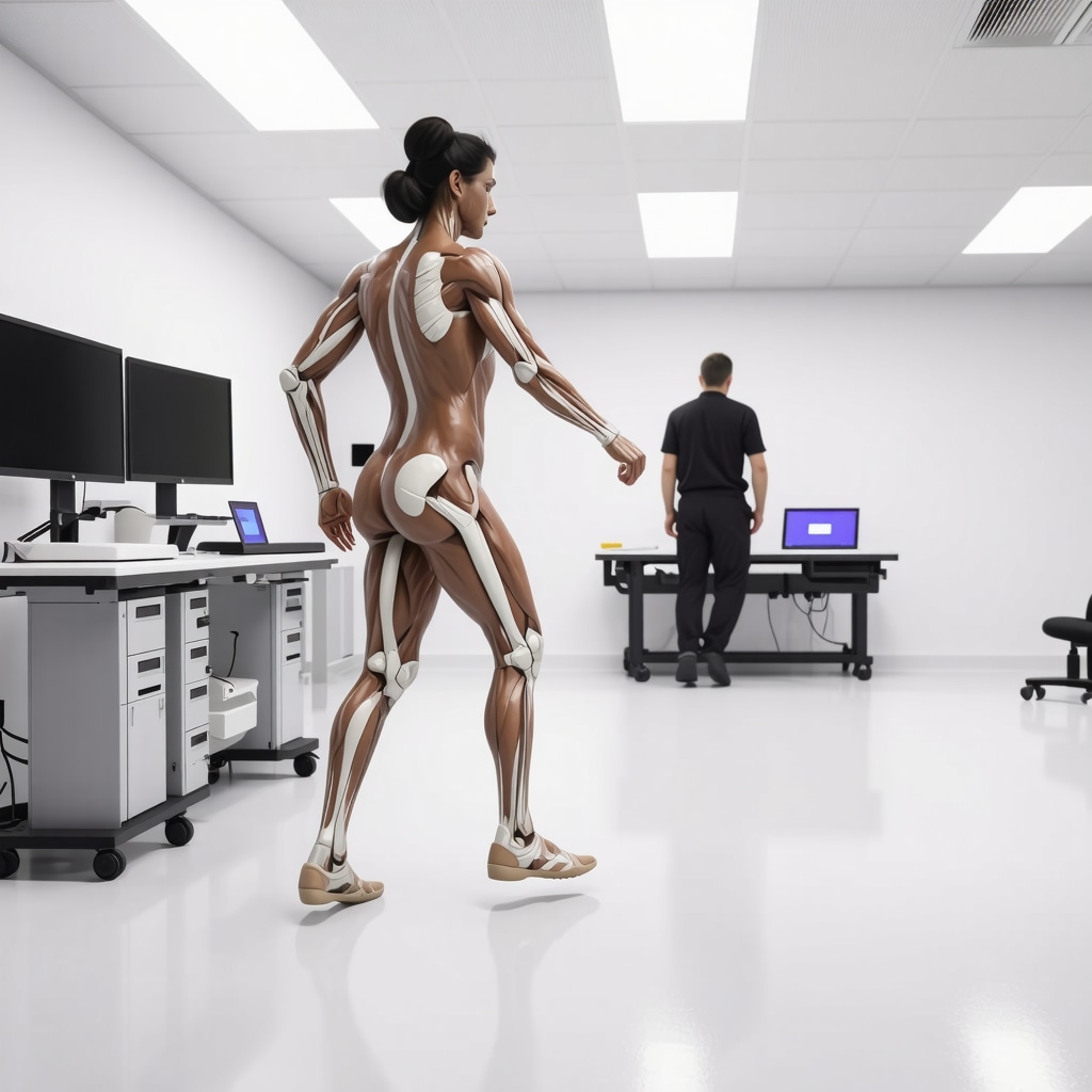
As orthopedic specialists push the boundaries of traditional assessment, biomechanical analysis emerges as a pivotal tool for understanding complex injury patterns. Modern gait analysis, force plate technology, and motion capture systems enable clinicians to quantify subtle deviations in movement that are often invisible during standard examinations. These insights are invaluable, especially in cases where repetitive strain or microtrauma contribute to chronic conditions. For example, a nuanced gait analysis can reveal asymmetric loading patterns that predispose workers to injury, allowing for targeted interventions that go beyond symptomatic relief.
How does biomechanical data enhance personalized treatment plans?
Biomechanical data provides a detailed map of functional deficits, informing tailored rehabilitation protocols. Studies published in the Journal of Orthopaedic Research (2022) underscore that integrating motion analysis with clinical assessments significantly improves outcomes in musculoskeletal disorder management. By combining subjective reports, physical exam findings, and objective biomechanical metrics, clinicians craft comprehensive strategies that address root causes rather than just symptoms.

The Role of Advanced Imaging Modalities in Detecting Hidden Pathologies
Beyond standard X-rays and MRIs, emerging imaging techniques like diffusion tensor imaging (DTI) and functional MRI (fMRI) are revolutionizing diagnostics. DTI, for instance, maps nerve fiber integrity with unprecedented precision, revealing microstructural damages that could explain persistent pain or neurological deficits post-injury. Such sophisticated imaging is especially relevant in complex cases involving nerve root impingement or subtle soft tissue injuries that traditional scans might miss.
Incorporating these modalities into evaluation protocols allows for early detection of pathologies that could otherwise remain undiagnosed until they cause significant disability. This proactive approach enables clinicians to initiate precise interventions, potentially reducing the duration and cost of treatment.
What are the implications of these advanced imaging techniques for future orthopedic practice?
As these technologies become more accessible and cost-effective, they promise to refine diagnostic accuracy further and facilitate personalized medicine. The integration of imaging biomarkers with clinical data could lead to predictive models for injury risk, thereby revolutionizing preventative strategies in occupational health.
For clinicians eager to adopt these innovations, collaboration with radiology specialists and ongoing education are vital. Staying abreast of the latest research ensures that patients benefit from cutting-edge diagnostics, ultimately translating into faster, more effective recovery pathways.
Harnessing Artificial Intelligence for Diagnostic Precision and Treatment Optimization
Perhaps the most exciting development in orthopedic evaluation is the application of artificial intelligence (AI). Machine learning algorithms analyze vast datasets—from imaging to patient histories—to identify patterns that escape human detection. AI-powered diagnostic tools can stratify injury severity, predict recovery timelines, and recommend personalized treatment plans with remarkable accuracy.
For instance, AI models trained on thousands of cases can flag subtle indicators of impending deterioration, prompting early intervention. This proactive stance not only improves patient outcomes but also optimizes resource allocation within healthcare systems.
Moreover, AI-driven virtual assistants support clinicians in decision-making processes, providing evidence-based suggestions tailored to individual patient profiles. As this technology matures, it promises a paradigm shift toward truly precision medicine in orthopedics.
How can healthcare providers effectively integrate AI into their orthopedic evaluation workflows?
Implementing AI requires careful planning—investing in robust data infrastructure, training staff, and maintaining ethical standards regarding data privacy. Engaging multidisciplinary teams, including data scientists and bioinformaticians, ensures that AI tools are both accurate and clinically relevant. As these systems evolve, continuous validation against real-world outcomes will be crucial to maintaining trust and efficacy.
To delve deeper into AI’s transformative potential, explore resources such as the American Academy of Orthopaedic Surgeons’ recent guidelines on digital health integration.
Embarking on this journey of advanced diagnostics and personalized care not only accelerates recovery but also elevates the standard of orthopedic practice. For practitioners committed to excellence, embracing these innovations is not just an option—they’re the future of effective, patient-centered injury management.
Why Do Subtle Diagnostic Clues Make All the Difference in Work Injury Assessments?
In the realm of orthopedic evaluations, especially for work-related injuries, the devil truly is in the details. Experienced clinicians have honed their ability to detect micro-signs—tiny deviations in gait, subtle neurological responses, or nuanced soft tissue signs—that can unveil underlying pathologies often missed during standard examinations. According to a study published in the Journal of Orthopaedic Research, these micro-criteria significantly enhance diagnostic accuracy, leading to tailored treatment plans that address root causes rather than just symptoms. For patients, this means faster recovery and reduced risk of chronic issues. This precision underscores the importance of specialized training and continuous education for orthopedic professionals, ensuring they remain adept at recognizing these crucial yet subtle clues.
How Does Biomechanical and Kinematic Analysis Elevate the Standard Orthopedic Evaluation?
Beyond traditional physical exams, integrating biomechanical and kinematic analysis provides a comprehensive view of a patient’s functional status. Technologies such as gait analysis systems, force plates, and motion capture allow clinicians to quantify movement deviations with remarkable precision. This data-driven approach reveals asymmetries, compensatory patterns, and micro-movements that predispose workers to injury or hinder recovery. A recent review in the Journal of Orthopaedic Research emphasizes that such detailed analysis informs personalized rehabilitation protocols, optimizing outcomes and preventing recurrence. For instance, identifying asymmetric loading during gait can lead to targeted orthotic interventions and specific strength training, dramatically improving recovery trajectories.
What Are the Emerging Frontiers in Imaging Technologies for Detecting Hidden Pathologies?
Innovations such as diffusion tensor imaging (DTI) and functional MRI (fMRI) are transforming diagnostic capabilities, especially for complex or elusive injuries. DTI’s ability to map nerve fiber integrity at microstructural levels enables clinicians to detect nerve damage that conventional MRI might miss, providing insights into persistent pain syndromes or subtle soft tissue injuries. A comprehensive review in the National Library of Medicine highlights these modalities’ potential to facilitate early, precise interventions, reducing the likelihood of long-term disability. As these technologies become more accessible, they promise a future where diagnostics are not only more accurate but also predictive—anticipating injuries before they fully manifest.
How Can Artificial Intelligence Revolutionize Orthopedic Evaluation and Patient Care?
Artificial intelligence (AI) stands poised to revolutionize orthopedic diagnostics by harnessing vast datasets—imaging, clinical histories, biomechanical metrics—and identifying complex patterns beyond human perception. Machine learning algorithms can stratify injury severity, forecast recovery timelines, and suggest personalized treatment pathways with unprecedented accuracy. For example, AI systems integrated into imaging platforms can flag subtle nerve impingements or soft tissue anomalies that might escape even seasoned clinicians. Studies in the Journal of Medical AI demonstrate that AI-powered decision support not only enhances diagnostic precision but also streamlines clinical workflows, reducing delays and improving patient outcomes. The challenge lies in thoughtful implementation—requiring collaboration across disciplines, rigorous validation, and adherence to ethical standards surrounding data privacy and bias mitigation.
What Steps Should Healthcare Providers Take to Integrate AI Effectively into Orthopedic Diagnostics?
Effective integration of AI entails investing in robust data infrastructure, training clinicians in AI literacy, and establishing multidisciplinary teams that include data scientists and radiologists. Ongoing validation studies are essential to ensure AI outputs remain accurate and clinically relevant. As the technology evolves, continuous feedback loops and real-world testing will be crucial to refining algorithms and building trust among users. By embracing these innovations, orthopedic practices can elevate their diagnostic accuracy, personalize treatment plans more effectively, and ultimately, accelerate patient recovery processes. For further insights into digital health adoption, explore the latest guidelines from the American Academy of Orthopaedic Surgeons.
Expert Insights & Advanced Considerations
1. Integration of Biomechanical Data Enhances Diagnostic Precision
Leveraging gait analysis, force plates, and motion capture systems allows clinicians to identify subtle movement deviations, leading to tailored treatment strategies that address root causes rather than just symptoms. This multidisciplinary approach improves recovery outcomes and minimizes recurrence risks.
2. Adoption of Cutting-Edge Imaging Modalities Transforms Detection of Hidden Pathologies
Emerging technologies such as diffusion tensor imaging (DTI) and functional MRI (fMRI) enable the visualization of microstructural nerve damage and soft tissue injuries. These tools facilitate early diagnosis and intervention, reducing long-term disability and enhancing patient prognosis.
3. Artificial Intelligence (AI) Revolutionizes Diagnostic and Treatment Processes
AI algorithms analyze vast datasets to predict injury severity, recovery timelines, and optimal treatment pathways. Integrating AI into clinical workflows supports personalized medicine, improves diagnostic accuracy, and streamlines decision-making, ultimately accelerating patient recovery.
4. Emphasis on Micro-Signs and Subtle Clues in Clinical Evaluation
Expert clinicians focus on micro-movements, neurological responses, and soft tissue signs that often escape standard exams. Recognizing these clues enhances diagnostic accuracy, enabling precise interventions that lead to faster and more effective recoveries.
5. Multidisciplinary Collaboration and Data Integration Are the Future
Combining insights from biomechanics, imaging, AI, and clinical examination fosters a comprehensive understanding of complex injuries. This integrated approach supports the development of personalized treatment plans that improve outcomes and patient satisfaction.
Curated Expert Resources
- Journal of Orthopaedic Research: Offers in-depth studies on biomechanical and kinematic analysis, emphasizing quantitative assessment techniques that advance diagnostic accuracy.
- National Library of Medicine (PubMed): Provides access to cutting-edge research articles on advanced imaging modalities like DTI and fMRI, highlighting their clinical applications in orthopedics.
- American Academy of Orthopaedic Surgeons (AAOS): Publishes guidelines and resources on integrating AI and digital health tools into orthopedic practice, fostering innovation and evidence-based care.
- Recent AI in Orthopedics Reports: Academic papers and industry reports on machine learning applications, predictive analytics, and decision support systems enhancing diagnosis and treatment.
Final Expert Perspective
In the evolving landscape of orthopedic injury evaluation, embracing advanced diagnostics such as biomechanical analysis, innovative imaging, and artificial intelligence is crucial for delivering top-tier patient care. These innovations empower clinicians to uncover subtle clues, diagnose complex conditions accurately, and craft personalized recovery plans. As you deepen your expertise, consider exploring the latest research and integrating multidisciplinary tools to stay at the forefront of orthopedic excellence. Your engagement and continuous learning are vital—share your insights or inquire about emerging technologies to contribute to a future where precision and personalization redefine recovery standards.
,
