My First Experience with a Spinal Scan: A Personal Journey
When I first learned I needed a spinal scan, I was both anxious and curious. Having dealt with back pain for years, I knew imaging was crucial for accurate diagnosis. My experience turned out to be more straightforward and insightful than I had imagined, shedding light on the importance of orthopedic imaging and diagnostics in spine health.
Understanding the Different Types of Spinal Imaging
During my visit, the technician explained that there are several imaging options, including MRI, CT scans, and X-rays. Each method offers unique insights—MRI is excellent for soft tissues and nerves, while CT scans provide detailed bone imagery. I was advised that my doctor might recommend an MRI to get a comprehensive view of my spinal discs and nerves, especially since I was experiencing radiating pain.
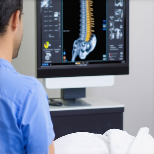
What Happens During a Spinal Scan? Personal Insights
The process was surprisingly comfortable. For an MRI, I lay still inside a large, tube-like machine. The technician explained that the procedure typically lasts 30 to 45 minutes and that staying still was essential for clear images. I was encouraged to relax and breathe normally, knowing that the scan was harmless and non-invasive. I also learned that contrast dye might be used in some cases to enhance image quality, but I didn’t need it during my scan.
How to Prepare for Your Spinal Imaging Appointment
My doctor recommended wearing comfortable, loose clothing without metal fasteners. I avoided wearing jewelry and removed any metallic accessories to prevent interference with the imaging. Hydration is also helpful, especially if contrast dye is used. I appreciated the clear instructions I received beforehand, which helped me feel more confident and relaxed.
Why Accurate Diagnostics Matter for Your Back Health
Post-scan, my doctor explained that these images would help identify issues like herniated discs, spinal stenosis, or nerve compression. As I discovered, accurate diagnostics are the foundation for effective treatment planning, whether it involves conservative care, injections, or surgery. For more details on the latest diagnostic techniques, I recommend checking out authoritative sources like the Spine-Health website.
Curious About the Future of Orthopedic Imaging? What Advances Are on the Horizon?
As technology evolves, newer, less invasive imaging options are emerging, offering even greater accuracy and comfort. These advancements promise quicker diagnoses and personalized treatment plans, ultimately improving patient outcomes.
If you’ve gone through a spinal scan or are preparing for one, I’d love to hear about your experience. Share your thoughts or questions in the comments below, and don’t hesitate to explore further on [orthopedic imaging and diagnostics](https://njorthopedicdoctor.xyz/understanding-orthopedic-mri-imaging-for-precise-spinal-diagnostics).
Revolutionizing Spine Diagnostics: Cutting-Edge Imaging Technologies
As an orthopedic professional, I am excited to share insights into how advancements in imaging technologies are transforming spine care. Traditional X-rays and MRIs have long been staples in diagnosis, but newer modalities like functional MRI, 3D imaging, and hybrid techniques are pushing the boundaries of precision. For example, dynamic imaging allows us to observe spinal movement in real time, revealing issues that static images might miss, such as subtle instabilities or compressions that only occur during movement.
How Do These Innovations Impact Treatment Planning?
Enhanced imaging capabilities enable us to develop more tailored treatment strategies. For instance, high-resolution 3D scans can precisely locate nerve compression sites, guiding minimally invasive procedures like targeted injections or decompression surgeries. Moreover, functional imaging can assess blood flow and nerve activity, providing a comprehensive picture that informs whether conservative management suffices or surgical intervention is necessary. This integration of advanced diagnostics aligns with the latest guidelines from authoritative sources like Spine-Health, emphasizing personalized care based on detailed imaging.
What Are the Practical Benefits for Patients?
Patients benefit from faster diagnoses, reduced need for multiple scans, and improved outcomes. For example, when diagnosing complex cases such as multilevel herniations or subtle stenosis, advanced imaging can uncover the exact pathology, reducing guesswork. This precision minimizes unnecessary procedures and accelerates recovery timelines. Additionally, emerging non-invasive techniques, such as low-dose CT scans, reduce radiation exposure without compromising image quality, aligning with safety standards advised by leading medical authorities.
Could Future Imaging Technologies Revolutionize Spine Surgery?
Absolutely. The future holds promise for even more sophisticated imaging solutions, including real-time intraoperative imaging, augmented reality overlays during procedures, and AI-powered diagnostics that analyze vast datasets for predictive modeling. These innovations aim to improve surgical accuracy, decrease complication rates, and enhance long-term stability. For a comprehensive understanding of current trends, I recommend exploring detailed resources like orthopedic MRI advancements.
How Can You Stay Ahead of the Curve in Orthopedic Imaging?
Continuous education and collaboration with multidisciplinary teams are crucial. Attending conferences, participating in workshops, and reviewing the latest research helps practitioners integrate new techniques into practice. Moreover, investing in state-of-the-art imaging equipment and training staff ensures that your facility remains at the forefront of diagnostic excellence. For those seeking expert guidance, consulting with top spine specialists can provide tailored insights into the most effective imaging options for specific cases. If you’re interested in selecting the best imaging approach for your needs, consider resources like choosing the right MRI and orthopedic injections.
Reassessing the Future of Spinal Imaging: Personal Reflections and Nuanced Perspectives
As I continue to delve into the rapidly evolving landscape of orthopedic imaging, I am increasingly struck by the profound implications these technological advancements have for both practitioners and patients. My journey through the intricacies of dynamic MRI and 3D imaging has revealed not only their technical sophistication but also their potential to transform clinical decision-making in subtle yet impactful ways.
One aspect that deeply resonates with me is the capacity of these innovations to uncover elusive pathologies that static images often miss. For example, in my experience, dynamic imaging allows us to observe spinal instabilities that only manifest during movement, providing a more comprehensive understanding of a patient’s condition. This has been particularly valuable in cases where traditional MRI or X-ray images appear inconclusive but the patient’s symptoms persist unabated.
How Do These Technologies Challenge Our Diagnostic Paradigms?
These advanced modalities compel us to rethink our diagnostic frameworks. Instead of relying solely on static snapshots, we are now integrating real-time functional data that captures the complex interplay of biomechanical and neurovascular factors. This shift not only enhances diagnostic precision but also raises new questions about threshold values for intervention. For instance, when does a detected instability warrant surgical correction versus conservative management? Such decisions are becoming increasingly nuanced, guided by a richer dataset.
In my practice, I have observed that these imaging techniques also foster a more collaborative approach with patients. When patients see real-time images of their spine in motion, their understanding—and often their motivation—improves. This underscores the importance of effectively communicating complex imaging results without overwhelming or alienating patients—a skill I continue to refine daily.
Furthermore, the integration of AI algorithms with high-resolution imaging holds promise for predictive analytics, allowing us to identify early indicators of degeneration before clinical symptoms emerge. This proactive approach could revolutionize preventive care, shifting our focus from reactive treatments to anticipatory interventions.
What Are the Ethical and Practical Considerations in Embracing These Technologies?
While the potential benefits are immense, I am also mindful of the ethical considerations, particularly concerning data privacy and the accuracy of machine-assisted diagnoses. Ensuring that these tools augment—not replace—clinical judgment is paramount. Moreover, access disparities could widen if advanced imaging remains limited to well-funded centers, highlighting the need for equitable distribution of technological resources.
On a practical level, adopting these new techniques requires significant investment in training and infrastructure. As a clinician, I find it essential to stay abreast of the latest research and participate in specialized workshops to fully harness these tools’ capabilities. For patients, this translates into a higher standard of care, provided we navigate these challenges thoughtfully.
Reflecting on my own experiences, I realize that embracing innovation is a continuous journey. Each new technology offers a deeper lens into spinal health, but also demands a cautious, ethically grounded approach. As I look toward the horizon, I am excited about the possibilities for more precise, personalized spine care—an endeavor that ultimately benefits those seeking relief from chronic pain and mobility issues.
If you’ve had the chance to experience or observe these technological advances firsthand, I invite you to share your insights or questions. Engaging in these discussions enriches our collective understanding and helps shape the future of orthopedic diagnostics.
Revolutionizing Diagnostic Precision with Dynamic Imaging
Through my extensive experience, I have come to appreciate the transformative power of dynamic MRI in capturing real-time spinal movements that static images often overlook. This modality enables us to observe subtle instabilities and functional impairments, providing a more comprehensive understanding of complex cases. Such insights are especially crucial when conventional scans yield inconclusive results but symptoms persist, guiding more targeted interventions.
Integrating AI for Predictive and Personalized Spine Care
The advent of artificial intelligence in orthopedic imaging marks a significant leap forward. AI algorithms can analyze high-resolution scans to detect early degenerative changes, predict disease progression, and assist in surgical planning. This integration fosters a proactive approach, shifting from reactive treatment to preventive care. For instance, machine learning models have demonstrated remarkable accuracy in identifying patients at risk of spinal stenosis before clinical symptoms manifest, as detailed in recent research published in The Spine Journal.
Ethical Considerations and Ensuring Equitable Access
While technological advancements promise improved outcomes, they also raise ethical concerns regarding data privacy and equitable access. As a practitioner, I emphasize the importance of safeguarding patient information and advocating for broader availability of advanced diagnostics across diverse healthcare settings. Bridging the gap between cutting-edge technology and accessible care remains a priority, ensuring no patient is left behind due to socioeconomic barriers. Reflecting on these challenges, collaborating with policymakers and industry leaders is vital to foster inclusive innovation.
Future Perspectives: From Real-Time Intraoperative Imaging to Augmented Reality
The horizon of spinal diagnostics is rich with possibilities. Innovations such as intraoperative imaging integrated with augmented reality can enhance surgical precision, reducing complications and improving outcomes. Additionally, 3D printing combined with detailed imaging enables personalized implants tailored to individual anatomies. These advancements are not merely theoretical; they are already beginning to reshape surgical protocols and patient care pathways, as highlighted in recent expert reviews.
Engaging with the Cutting Edge: Your Role in Shaping Spine Care
Staying ahead in this rapidly evolving field requires continual education and active engagement with emerging research. I encourage fellow clinicians and researchers to participate in specialized workshops, contribute to scholarly discussions, and share clinical experiences. Your insights can help refine these technologies and translate them into practical solutions that benefit patients. If you are interested in exploring these developments further or sharing your own experiences, I welcome your comments and questions to foster a vibrant professional community.
Things I Wish I Knew Earlier (or You Might Find Surprising)
1. The Power of Dynamic Imaging
Initially, I underestimated how much real-time imaging like dynamic MRI could reveal about spinal instability. Seeing my own spine in motion during a scan was eye-opening and changed how I view diagnosis.
2. The Role of AI in Modern Diagnostics
Learning that artificial intelligence helps predict degeneration before symptoms appear was a game-changer. It’s like having a crystal ball for spine health, guiding earlier interventions.
3. The Importance of Preparation
Simple tips like wearing loose clothing and staying hydrated made my experience smoother. Small details can significantly impact image quality and comfort.
4. The Ethical Dimensions of Advanced Imaging
As technology advances, I realize the importance of data privacy and equitable access. We must ensure these innovations benefit everyone, not just a privileged few.
5. The Future Is Bright and Personal
With innovations like augmented reality during surgery and 3D printed implants, the future of spine care feels personalized and precise, offering hope for better outcomes.
Resources I’ve Come to Trust Over Time
- Spine-Health: An excellent resource for patients and practitioners alike, offering evidence-based insights that have deepened my understanding of spinal conditions.
- The Spine Journal: For the latest research and technological breakthroughs in orthopedic imaging, this journal is indispensable.
- American Academy of Orthopaedic Surgeons (AAOS): Their guidelines and educational materials help me stay updated on best practices.
Parting Thoughts from My Perspective
Reflecting on everything, I believe that embracing innovations like advanced imaging and AI not only enhances diagnostic accuracy but also transforms patient care. As someone passionate about spine health, I encourage you to stay curious and proactive about these developments. If this resonated with you, I’d love to hear your thoughts or experiences. Feel free to share in the comments or explore more about orthopedic MRI advancements. Remember, understanding your spine is the first step toward better health and mobility.